. 2010 Jul 30;183(1):21-9.
doi: 10.1016/j.pscychresns.2010.04.016. Epub 2010 Jun 9.

White matter volume abnormalities and associations with symptomatology in schizophrenia

Affiliations

White matter volume abnormalities and associations with symptomatology in schizophrenia

Nikolaos Makris et al. Psychiatry Res. .

Abstract

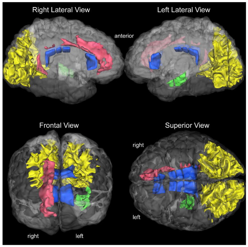
The cerebral white matter (WM) is critically involved in many bio-behavioral functions impaired in schizophrenia. However, the specific neural systems underlying symptomatology in schizophrenia are not well known. By comparing the volume of all brain fiber systems between chronic patients with DSM-III-R schizophrenia (n=88) and matched healthy community controls (n=40), we found that a set of a priori WM regions of local and distal associative fiber systems was significantly different in patients with schizophrenia. There were significant positive correlations between volumes (larger) in anterior callosal, cingulate and temporal deep WM regions (related to distal connections) with positive symptoms, such as hallucinations, delusions and bizarre behavior, and significant negative correlation between volumes (smaller) in occipital and paralimbic superficial WM (related to local connections) and posterior callosal fiber systems with higher negative symptoms, such as alogia. Furthermore, the temporal sagittal system showed significant rightward asymmetry between patients and controls. These observations suggest a pattern of volume WM alterations associated with symptomatology in schizophrenia that may be related in part to predisposition to schizophrenia.


Figures

Figure 1
Figure 1. Segmentation and Cortical and White Matter Parcellation
In a-c, the raw image of a coronal section (a) and the Center for Morphometric Analysis (CMA) general segmentation methodology of the cortical and subcortical structures (b, c) is shown (Filipek et al., 1994; Makris et al., 1999). The result of the detailed CMA cortical parcellation is shown in d (Rademacher et al., 1992; Caviness et al., 1996b). In e, the result of the CMA white matter parcellation technique used in the present study (Makris et al., 1999; Meyer et al., 1999) is shown.
Figure 2
Figure 2. Topographic and Volumetric White Matter Parcellation
CMA white matter parcellation follows general segmentation and cortical parcellation and is a virtually an automated procedure, which requires a manual pre-processing step consisting on the identification and selection of six anatomical nodal points at the corpus callosum and deep gray matter (Makris et al., 1999; Meyer et al., 1999). Although minimal in terms of time required, this manual step is critical to guarantee the anatomic accuracy of the procedure. This anatomically-specified system of analysis allows parcellation of the cerebral white matter into a superficial zone or superficial white matter (referred in this figure as “radiate” zone) and a deep zone, which contains the sagittal strata systems or sagittal systems and the bridging systems (referred in this figure as “deep and bridging” zone). The sagittal systems include three major classes of axonal systems: a) the major cortico-cortical ipsilateral long association fiber pathways, b) the projection fibers linking cortex, thalamus, basal ganglia, amygdala, hippocampus, pons, brainstem, and spinal cord, and c) the callosal commissural system. The bridging systems consist of the internal capsule, basal forebrain fiber systems such as the ventral amygdalofugal pathway, as well as the fornix, the anterior commissure and the dorsal and ventral hippocampal commissures (Makris et al., 1999). The parcellation of the radiate zone provides 47 parcellation units of superficial white matter shown in a color-coded scheme in the plate on the far left side of this figure.
Figure 3
Figure 3. White Matter Abnormalities in the Cerebrum in Schizophrenia
The salient results of this study are shown in a series of 3-D visualizations in a human cerebrum. The white matter regions abnormal in schizophrenia are rendered in a color-coded fashion. The occipital superficial white matter bilaterally is shown in yellow, right paralimbic white matter (including the cingulum) shown in red, the left deep temporal sagittal system white matter shown in green, and the majority of the corpus callosum shown in dark blue. The 3-D reconstructions have been done using an in-house tool called SVV (surface and volume visualizer).

Similar articles

Cited by

References

    1. Andreasen NC, Arndt S, Swayze V, 2nd, Cizaldo T, Flaum M, O'Leary D, et al. Thalamic abnormalities in schizophrenia visualized through magnetic resonance image averaging. Science. 1994;266(5183):294–298. - PubMed
    1. Andreasen NC, Nopoulos P, O'Leary DS, Miller DD, Wassink T, Flaum M. Defining the phenotype of schizophrenia: cognitive dysmetria and its neural mechanisms. Biological Psychiatry. 1999;46(7):908–920. - PubMed
    1. Andreasen NC, Olsen S. Negative versus positive schizophrenia. Definition and validation. Archives of General Psychiatry. 1982;39:789–794. - PubMed
    1. Annett M. A classification of hand preference by association analysis. British Journal of Psychology. 1970;61(3):303–321. - PubMed
    1. Argartz I, Andersson J, Skare S. Abnormal brain white matter in schizophrenia: A diffusion tensor imaging study. Neuroreport. 2001;12(10):2251–2254. - PubMed

Publication types