. 2010 Aug 13;285(33):25753-66.
doi: 10.1074/jbc.M109.093468. Epub 2010 Jun 10.

Autoactivation of transforming growth factor beta-activated kinase 1 is a sequential bimolecular process

Affiliations

Autoactivation of transforming growth factor beta-activated kinase 1 is a sequential bimolecular process

Roland Scholz et al. J Biol Chem. .

Abstract

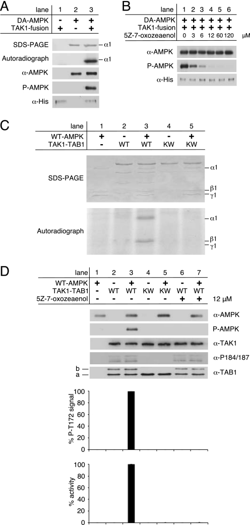
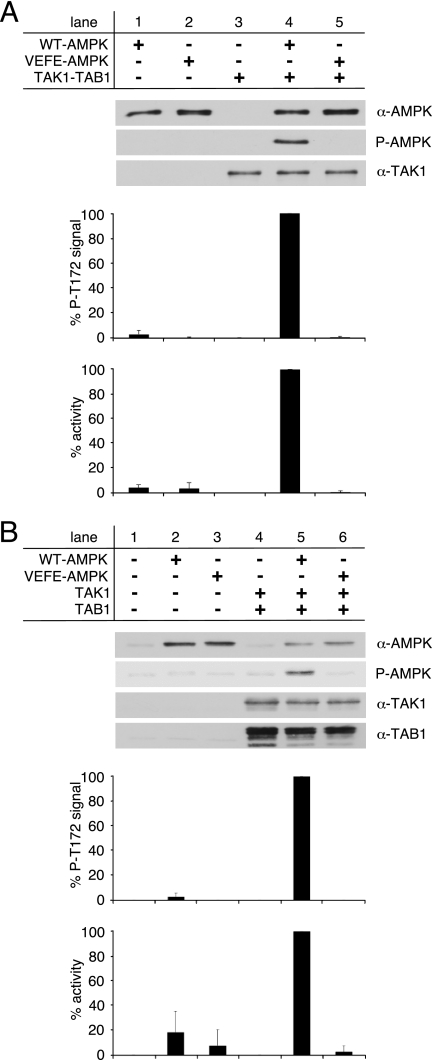
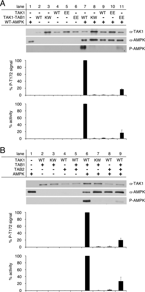
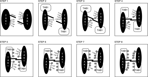
Transforming growth factor-beta-activated kinase 1 (TAK1), an MAP3K, is a key player in processing a multitude of inflammatory stimuli. TAK1 autoactivation involves the interplay with TAK1-binding proteins (TAB), e.g. TAB1 and TAB2, and phosphorylation of several activation segment residues. However, the TAK1 autoactivation is not yet fully understood on the molecular level due to the static nature of available x-ray structural data and the complexity of cellular systems applied for investigation. Here, we established a bacterial expression system to generate recombinant mammalian TAK1 complexes. Co-expression of TAK1 and TAB1, but not TAB2, resulted in a functional and active TAK1-TAB1 complex capable of directly activating full-length heterotrimeric mammalian AMP-activated protein kinase (AMPK) in vitro. TAK1-dependent AMPK activation was mediated via hydrophobic residues of the AMPK kinase domain alphaG-helix as observed in vitro and in transfected cell culture. Co-immunoprecipitation of differently epitope-tagged TAK1 from transfected cells and mutation of hydrophobic alphaG-helix residues in TAK1 point to an intermolecular mechanism of TAB1-induced TAK1 autoactivation, as TAK1 autophosphorylation of the activation segment was impaired in these mutants. TAB1 phosphorylation was enhanced in a subset of these mutants, indicating a critical role of alphaG-helix residues in this process. Analyses of phosphorylation site mutants of the activation segment indicate that autophosphorylation of Ser-192 precedes TAB1 phosphorylation and is followed by sequential phosphorylation of Thr-178, Thr-187, and finally Thr-184. Finally, we present a model for the chronological order of events governing TAB1-induced TAK1 autoactivation.


Figures

FIGURE 1.
FIGURE 1.
Analysis of TAK1 containing complexes after two-dimensional purification. A, Western blot analysis of bacterially expressed His-TAK1 containing complexes subsequent to two-dimensional purification. Anti-TAK1, anti-TAB1, anti-TAB2, and phospho-specific anti-TAK1-Thr(P)-184 (α-P184), anti-TAK1-Thr-187 (α-P187), and anti-TAK1-Thr(P)-184/187 (α-P184/187) antibodies were used for detection. KW-TAK1 represents the kinase-deficient mutant His-TAK1(K63W). For better comparability of activation segment phosphorylation and protein complex composition, the amount of analyzed protein was adjusted to equal His-TAK1 content. B, Western blot analysis of bacterially expressed and purified His-TAK1-TAB1 subjected to size exclusion chromatography using antibodies as in A. 1-ml fractions were collected, and every third fraction was analyzed. The corresponding elution volume V(Elu) is specified. The given molecular weights correspond to the electrophoretic mobility estimated from SDS-PAGE. Line a, unmodified TAB1 species; line b, shifted TAB1 species.
FIGURE 2.
FIGURE 2.
Activation of AMPK by active TAK1 in vitro. A, AMPK kinase-dead mutant α1(D157A)β1γ1 (DA-AMPK) was incubated with commercially available, active His-tagged TAK1-TAB1 fusion protein from baculoviral expression (TAK1 fusion) in the presence of [γ-32P]ATP for 20 min at 37 °C. Phosphorylation of DA-AMPK was detected by incorporation of 32P followed by autoradiography and Western blot analysis using anti-AMPKα and phospho-specific anti-AMPKα Thr-172 antibodies (P-AMPK). The input of TAK1-fusion protein was probed with anti-His antibody. B, 25 ng of TAK1 fusion was preincubated with increasing concentrations of the TAK1-selective inhibitor (5Z)-7-oxozeaenol in 10 mm Hepes, pH 7.5, for 15 min at 37 °C before adding DA-AMPK. C, phosphorylation of recombinant WT-AMPK by the bacterially expressed recombinant wild-type TAK1-TAB1 (WT) and the kinase-deficient mutant TAK1(K63W)-TAB1 (KW) in the presence of [γ-32P]ATP was monitored by autoradiography. The corresponding Coomassie Blue-stained SDS-polyacrylamide gel is shown on the top. D, in vitro activation of recombinant WT-AMPK by bacterially expressed and purified recombinant WT-TAK1-TAB1 and KW-TAK1-TAB1. TAK1-TAB1 proteins were preincubated for 15 min at 37 °C in the presence or absence of 12 μm (5Z)-7-oxozeaenol. Western blot analysis was performed with anti-AMPKα, anti-TAK1, anti-TAB1, and phospho-specific anti-AMPKα Thr-172 (P-AMPK) and anti-TAK1 Thr(P)-184/187 (α-P184/187) antibodies. To quantify AMPKα Thr-172 phosphorylation as a consequence of TAK1 activity, the respective background signals of WT-AMPK (lane 1) and TAK1-TAB1 complexes (lanes 2 and 4) were subtracted, and the AMPKα phospho-Thr172 signals were normalized to AMPKα and TAK1 signal intensities. AMPK activity was determined, and respective background activities were subtracted. All activities were standardized to corresponding TAK1 signal intensities as revealed from densitometric Western blot analysis to determine TAK1 activity in relation to TAK1 amount. All data were normalized relative to WT-AMPK activation by WT-TAK1-TAB1 (n = 3; S.D.). Line a, unmodified TAB1 species; line b, shifted TAB1 species.
FIGURE 3.
FIGURE 3.
Activation of WT- and VEFE-AMPK by active TAK1. A, in vitro activation of recombinant WT- and VEFE-AMPK by bacterially expressed and purified TAK1-TAB1. Western blot analysis was performed using anti-TAK1, anti-AMPKα, and phospho-specific anti-AMPKα Thr-172 (P-AMPK) antibodies. TAK1 background signal (lane 3) was subtracted, and AMPKα phospho-Thr-172 signal intensities were standardized to AMPKα signals. Activity was determined by a HPLC- and SAMS-peptide-based activity assay. Background signal of TAK1 was subtracted, and data were normalized to WT-AMPK activation by TAK1-TAB1 (n = 3; S.D.). B, HeLa cells were transfected with plasmids encoding Myc-AMPKα1 wild-type and VEFE mutant, FLAG-TAK1, and HA-TAB1. Cells were treated with 1 mm hydrogen peroxide for 15 min before lysis. HeLa cell lysates were subjected to Western blot analysis using anti-TAK1, anti-TAB1, anti-AMPKα, and phospho-specific anti-AMPKα Thr-172 (P-AMPK) antibodies. Subsequent to immunoprecipitation using anti-AMPKα1 antibodies, AMPK activity was determined. Respective background signals from mock- and TAK1/TAB1-double transfected cells were subtracted, and data were normalized to the WT-AMPK/TAK1/TAB1 triple-transfected cells (n = 3; S.D.).
FIGURE 4.
FIGURE 4.
Activation of TAK1 requires co-expression of TAB1. A, recombinant WT-AMPK was activated by bacterially expressed TAK1 and TAK1 co-expressed with TAB1 (TAK1-TAB1). Here, the activities of wild-type TAK1 proteins were additionally compared with the activities of TAK1 proteins carrying the phosphorylation site mimicking mutation T178E,T184E (EE) within the TAK1 activation segment. B, activity of recombinant TAK1 bacterially co-expressed with TAB1 and/or TAB2. The kinase-deficient mutant TAK1(K63W)-TAB1 (KW) served as a control. Western blot analysis utilized anti-TAK1, anti-AMPKα, and phospho-specific anti-AMPKα Thr-172 (P-AMPK) antibodies. The AMPKα phospho-Thr-172 signals were quantified after subtraction of respective WT-AMPK (lane 1) and TAK1 background signals (lanes 2–6) and normalization to AMPKα and TAK1 signals. Activity was determined, and respective background activities were subtracted. All activities were standardized to corresponding TAK1 signal intensities as revealed from densitometric Western blot analysis. The data were normalized relative to WT-AMPK activation by WT-TAK1-TAB1 (n = 3; S.D.).
FIGURE 5.
FIGURE 5.
Involvement of TAK1 αG-helix residues in TAB1 and intermolecular TAK1 phosphorylation. A, AMPKα subunit double knock-out (α1−/−, α2−/−) MEFs were transfected with plasmids encoding HA-TAK1 and FLAG-TAK1. Immunoprecipitation (IP) from cell lysates was performed using an anti-HA antibody. Immunoprecipitates were analyzed by Western blot with anti-FLAG, anti-HA, and anti-TAK1 antibodies. B, six times excess of bacterially expressed kinase-deficient KW-TAK1-TAB1 (150 μg/ml) was incubated with WT-TAK1-TAB1 (25 μg/ml). Intermolecular TAK1 activation segment phosphorylation was monitored by Western blot analysis using anti-TAK1, anti-TAB1, and phospho-specific anti-TAK1-Thr(P)-184/187 (α-P184/187) antibodies. 175 μg/ml KW-TAK1-TAB1 and WT-TAK1-TAB1 were used as a control. C, alignment of the αG-helix region sequences of Snf1 from Saccharomyces cerevisiae, AMPK α1 and α2 subunits, and CamKK2 from Rattus norvegicus, LKB1 from Mus musculus, and TAK1 from human (GenBankTM accession numbers P06782, AAC52355, CAA82620, NP_112628, NP_035622, and O43318). Residues highlighted in black show conservation in all sequences, whereas residues highlighted in gray show partial conservation. The position of the hydrophobic AMPK kinase domain αG-helix residues Val-219 and Phe-223 is specified. Hydrophobic TAK1 kinase domain αG-helix residues Ala-236, Phe-237, Ile-239, Met-240, Trp-241, and Val-243 are boxed in red. D, structural analysis of the TAK1 kinase domain (Protein Data Bank code 2eva). Surface-exposed location of the αG-helix with hydrophobic residues highlighted in red (hydrophobicity scale >0.2 (64)) and rather hydrophilic residues highlighted in blue (hydrophobicity scale < 0.0 (64)) is shown. Structurally unresolved activation segment residues 179–190 are represented by a dashed line. Activation segment phosphorylation sites Ser-192 and Thr-178 are shown in green. E, upper panel, Western blot analysis of bacterially expressed WT- and KW-TAK1-TAB1 as well as TAK1-TAB1 mutants with hydrophobic αG-helix residues Ala-236, Phe-237, Ile-239, Met-240, Trp-241, and Val-243 replaced by glutamate-TAK1(A236E, M240E)-TAB1 (AM), TAK1(F237E, W241E)-TAB1 (FW), TAK1(W241E, V243E)-TAB1 (WV), and TAK1(I239E, V243E)-TAB1 (IV). Lower panel as in upper panel, but the corresponding αG-helix residues are substituted by lysine instead of glutamate. Line a, unmodified TAB1 species; line b, shifted TAB1 species.
FIGURE 6.
FIGURE 6.
Sequence of phosphorylation events within the TAK1 kinase domain activation segment. Western blot analysis of bacterially expressed WT-TAK1-TAB1, KW-TAK1-TAB1 TAK1-TAB1 mutants carrying single amino acid mutations at phosphorylation sites within the TAK1 kinase domain activation segment: TAK1(T178A)-TAB1, TAK1(T178E)-TAB1, TAK1(T184A)-TAB1, TAK1(T184E)-TAB1, TAK1(T187A)-TAB1, TAK1(T187E)-TAB1, TAK1(S192A)-TAB1, and TAK1(S192E)-TAB1. Anti-TAK1, anti-TAB1, and phospho-specific anti-TAK1-Thr(P)-184 (α-P184), anti-TAK1-Thr(P)-187 (α-P187), and anti-TAK1-Thr(P)-184/187 (α-P184/187) antibodies were applied. Line a, unmodified TAB1 species; line b, shifted TAB1 species.
FIGURE 7.
FIGURE 7.
Activity of TAK1-TAB1 activation segment mutants. A, in vitro activation of recombinant WT-AMPK by bacterially expressed WT- and KW-TAK1-TAB1 as well as TAK1(T178A)-TAB1 and TAK1(T178E)-TAB1. B–D as in A but analyzing TAK1(T184A)-TAB1 and TAK1(T184E)-TAB1, TAK1(T187A)-TAB1 and TAK1(T187E)-TAB1, and TAK1(S192A)-TAB1 and TAK1(S192E)-TAB1, respectively. Western blot analysis was performed using anti-TAK1, anti-AMPKα, and phospho-specific anti-AMPKα Thr-172 (P-AMPK) antibodies. To quantify AMPKα Thr-172 phosphorylation, the respective background signals from nonactivated AMPK (lane 1) and TAK1-TAB1 (lane 2–5) were subtracted, and phospho-Thr-172 signal intensities were normalized to AMPKα and TAK1 signals. Activity was determined, and respective background activities (lane 1–5) were subtracted. All activities were standardized to TAK1 signal intensities as revealed from densitometric Western blot analysis. Data were normalized relative to WT-AMPK activation by WT-TAK1-TAB1 (n = 3; S.D.).
FIGURE 8.
FIGURE 8.
Model of sequential TAB1-induced TAK1 autoactivation. Step 1, TAK1 activation segment autophosphorylation sites Thr-178, Thr-184, Thr-187, and Ser-192 are not accessible in the absence of TAB1. Step 2, upon TAB1 binding to the TAK1 catalytic domain, a conformational change is induced in TAK1, leading to improved accessibility of Ser-192. Step 3, Ser-192 autophosphorylation is mediated by an intermolecular mechanism, which involves hydrophobic residues of the TAK1 kinase domain αG-helix. Step 4, Ser-192 phosphorylation re-positions the αG-helix, thus facilitating the further functional interplay between TAB1 and TAK1. Step 5, TAB1 gets phosphorylated, which results in improved accessibility of the remaining activation segment autophosphorylation sites Thr-178, Thr-184, and Thr-187. Steps 6–8, completion of TAB1-induced TAK1 autoactivation by αG-helix-mediated sequential intermolecular autophosphorylation of Thr-178, Thr-187, and finally Thr-184.

Similar articles

Cited by

References

    1. Yamaguchi K., Shirakabe K., Shibuya H., Irie K., Oishi I., Ueno N., Taniguchi T., Nishida E., Matsumoto K. (1995) Science 270, 2008–2011 - PubMed
    1. Ninomiya-Tsuji J., Kishimoto K., Hiyama A., Inoue J., Cao Z., Matsumoto K. (1999) Nature 398, 252–256 - PubMed
    1. Wang C., Deng L., Hong M., Akkaraju G. R., Inoue J., Chen Z. J. (2001) Nature 412, 346–351 - PubMed
    1. Takaesu G., Surabhi R. M., Park K. J., Ninomiya-Tsuji J., Matsumoto K., Gaynor R. B. (2003) J. Mol. Biol. 326, 105–115 - PubMed
    1. Lee J., Mira-Arbibe L., Ulevitch R. J. (2000) J. Leukocyte Biol. 68, 909–915 - PubMed

Publication types

MeSH terms

Substances