. 2010 Aug 13;285(33):25296-307.
doi: 10.1074/jbc.M110.128801. Epub 2010 Jun 9.

In silico docking and electrophysiological characterization of lacosamide binding sites on collapsin response mediator protein-2 identifies a pocket important in modulating sodium channel slow inactivation

Affiliations

In silico docking and electrophysiological characterization of lacosamide binding sites on collapsin response mediator protein-2 identifies a pocket important in modulating sodium channel slow inactivation

Yuying Wang et al. J Biol Chem. .

Abstract

The anti-epileptic drug (R)-lacosamide ((2R)-2-(acetylamino)-N-benzyl-3-methoxypropanamide (LCM)) modulates voltage-gated sodium channels (VGSCs) by preferentially interacting with slow inactivated sodium channels, but the observation that LCM binds to collapsin response mediator protein 2 (CRMP-2) suggests additional mechanisms of action for LCM. We postulated that CRMP-2 levels affects the actions of LCM on VGSCs. CRMP-2 labeling by LCM analogs was competitively displaced by excess LCM in rat brain lysates. Manipulation of CRMP-2 levels in the neuronal model system CAD cells affected slow inactivation of VGSCs without any effects on other voltage-dependent properties. In silico docking was performed to identify putative binding sites in CRMP-2 that may modulate the effects of LCM on VGSCs. These studies identified five cavities in CRMP-2 that can accommodate LCM. CRMP-2 alanine mutants of key residues within these cavities were functionally similar to wild-type CRMP-2 as assessed by similar levels of enhancement in dendritic complexity of cortical neurons. Next, we examined the effects of expression of wild-type and mutant CRMP-2 constructs on voltage-sensitive properties of VGSCs in CAD cells: 1) steady-state voltage-dependent activation and fast-inactivation properties were not affected by LCM, 2) CRMP-2 single alanine mutants reduced the LCM-mediated effects on the ability of endogenous Na(+) channels to transition to a slow inactivated state, and 3) a quintuplicate CRMP-2 alanine mutant further decreased this slow inactivated fraction. Collectively, these results identify key CRMP-2 residues that can coordinate LCM binding thus making it more effective on its primary clinical target.


Figures

FIGURE 1.
FIGURE 1.
(R)-Lacosamide binds to CRMP-2 in rat brain lysates. A, in vitro labeling of endogenous CRMP-2 in rat brain lysate. Rat brain lysate (∼110 μg) was incubated with the affinity bait isothiocyanate and chemical reporter (AB-NCS&CR; 10 μm) LCM for 20 min at room temperature in the absence or presence of increasing amounts of LCM with 500 equivalent (eq) amounting to 5 mm LCM. AB&CR agent-labeled proteins were detected by in-gel fluorescence scanning after Cu(I)-mediated cycloaddition to a rhodamine-azide reporter probe. An inverted black and white image is presented. B, CRMP-2 protein was detected by Western blot analysis using an anti-CRMP-2 antibody. C, a gray scale image of the Coomassie Blue-stained gel in A showing equal loading of proteins in the various samples. Representative results from a total of three experiments are shown. Molecular mass markers are indicated in kilodaltons.
FIGURE 2.
FIGURE 2.
Enantioselective modification of CRMP-2 by LCM affects steady-state slow inactivation state of Na+ currents in CAD cells. A, currents were evoked by 5-s prepulses between −120 mV and −20 mV, and then fast inactivated channels were allowed to recover for 150 ms at a hyperpolarized pulse to −120 mV. The fraction of channels available at 0 mV was analyzed. Representative current traces are from a CRMP-2-EGFP-overexpressing CAD cells in the absence (left traces) or presence (right traces) of LCM treatment. The black trace in each panel represents the current at −50 mV. B, summary of steady-state slow activation curves for CAD cells transfected with CRMP-2-EGFP before and after application of (R)- or (S)-LCM (100 μm). CRMP-2 overexpression blocked the development of LCM-induced slow inactivation, which was observed in EGFP-overexpressing neurons treated with LCM. C, summary of the fraction of current available at −50 mV for CRMP-2-expressing CAD cells in the absence or presence of (R)- or (S)-LCM (100 μm). (R)-, but not (S)-, LCM significantly reduced the fraction of current available at −50 mV (*, p < 0. 05, Student's t test). Data are from 7–9 cells per condition. D, Western blot analysis with a CRMP-2 antibody showing successful (>90%) knockdown of CRMP-2 protein in CAD cells transfected with CRMP-2 siRNA compared with those transfected with scramble siRNA for 2 days. Under the same conditions, the expression the β-tubulin protein was unchanged. Duplicate lanes are from separate experiments.
FIGURE 3.
FIGURE 3.
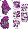
In silico docking identifies five putative (R)-lacosamide binding pockets on CRMP-2. A and B, surface representations of the three-dimensional structure of the CRMP-2 monomer (PDB code: 2GSE) with the locations of the putative binding pockets (1–5) highlighted in green. One or more LCMs bound to the five binding cavities within CRMP-2 are indicated. LCM is shown in capped-sticks representation. LCM is color-coded according to atom types (C, N, and O in white, blue, and red, respectively). The structure in B is rotated ∼180° relative to that in A. C, an enlarged view of each of the five binding pockets. C atoms are shown in yellow. Only the predominant conformational state of LCM is shown in each pocket. The asterisk denotes the position of amino acid Ser-421 (orange residue) that is adjacent, but not predicted to be within coordinating range, of putative binding pocket 2. The residues coordinating LCM binding are indicated in single amino acid letter code. Residues in bold (shown in blue) indicate positions that were mutated to alanine. Hydrogen bonds are shown with dashed lines.
FIGURE 4.
FIGURE 4.
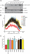
Expression and functional characterization of lacosamide binding CRMP-2 alanine mutants in embryonic cortical neurons. A, total protein lysates (20 μg each) from CAD cells expressing EGFP, CRMP-2-EGFP, or the various mutants were immunoblotted with antibodies against EGFP (i and ii), CRMP-2 (iii), and β-tubulin (iv). All CRMP-2-EGFP alanine mutants expressed well in CAD cells. Representative blots from three separate experiments are shown. Molecular mass standards are indicated on the left (kilodaltons). B, dendritic branching was calculated by Sholl analysis (41), which measures the number of neurites crossing concentric circles (intersections or branch points) at various radial distances from the cell soma. Sholl analysis of low density cultures of cortical neurons (n = 10–12 per condition) transfected at 3 DIV (and grown for 48 h) revealed a significant increase in the number of processes, reaching a peak of ∼20 crossings at ∼75–100 μm from the soma in wild-type and mutant CRMP-2-EGFP-transfected cells compared with EGFP transfected cells which reached a peak crossing of 8. C, summary of the average peak dendritic complexity for EGFP, wild-type CRMP-2EGFP, and mutant CRMP-2-EGFP transfected cortical neurons. A significant increase in neuritic complexity was seen in wild-type and all mutant CRMP-2-EGFP compared with EGFP-expressing (*, p < 0.05 versus EGFP at each distance between ∼60 and 120 μm; Student's t test).
FIGURE 5.
FIGURE 5.
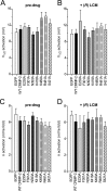
Activation properties of Na+ currents are not affected by LCM in CAD cells expressing wild-type or mutant CRMP-2 proteins. Values for V½, the voltage of half-maximal activation and the slope factors (k) were derived from Boltzmann distribution fits to the individual recordings and averaged to determine the mean (±S.E.) voltage dependence of activation. The V½ (A and B) and k (C and D) of activation were not different between any of the conditions tested and irrespective of drug treatment (p > 0.05, one-way ANOVA).
FIGURE 6.
FIGURE 6.
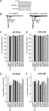
Fast inactivation properties of Na+ currents are not affected by LCM in CAD cells expressing wild-type or mutant CRMP-2 proteins. A, voltage protocol for fast inactivation. B, representative current traces showing voltage-dependent fast inactivation of sodium currents from CAD cells expressing wild-type CRMP-2 in the absence (left) or presence (right) of 100 μm LCM. Values for V½, the voltage of half-maximal inactivation, and the slope factors (k) were derived from Boltzmann distribution fits to the individual recordings and averaged to determine the mean (±S.E.) voltage dependence of steady-state inactivation. The V½ (C and D) and k (E and F) of steady-state fast inactivation were not different between any of the conditions tested and irrespective of drug treatment (p > 0.05, one-way ANOVA).
FIGURE 7.
FIGURE 7.
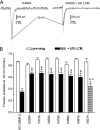
(R)-Lacosamide increases the fraction of Na+ current available in CAD cells expressing CRMP-2 mutants. A, representative traces from CAD cells expressing K480A CRMP-2-EGFP in response to a voltage protocol (see inset in Fig. 2A) designed to measure slow inactivation. Current traces in the absence (left traces) or presence (right traces) of 100 μm LCM treatment are shown. The black trace in each panel represents the current at −50 mV. Traces are shown for the K480A only, because all other mutants behaved in a similar manner. B, summary of the average fraction of current available, at −50 mV, in the absence (open bars) and presence (filled bars) of LCM (n = 7–9 cells per condition). LCM decreased the fraction of current available compared with pre-drug values for wild-type (WT) and mutant CRMP-2 proteins (*, p < 0.05, ANOVA with Dunnett's post-hoc test). The fraction of current available in CAD cells expressing the various CRMP-2 mutants in the presence of LCM was significantly greater than that in wild-type CRMP-2 (&, p < 0.05, ANOVA with Dunnett's post-hoc test). In contrast, the fraction of current available in CAD cells expressing the S421A CRMP-2 mutant in the presence of LCM was not significantly greater than that in wild-type CRMP-2 (a, p = 0.0895) but was significantly smaller than any of the other CRMP-2 mutants (b, p < 0.05, ANOVA with Dunnett's post-hoc test).
FIGURE 8.
FIGURE 8.
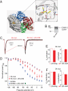
A quintuplet alanine mutant of CRMP-2 reduces (R)-lacosamide-induced shift in slow inactivation of Na+ currents in CAD cells. A, three-dimensional structure of the CRMP-2 tetramer (PDB code: 2GSE). Each subunit within the tetramer structure is shown in a different color. The subunit used for the docking is shown in gray surface representation, whereas the remaining three are depicted in blue, red, and green ribbon representation, respectively. LCM is shown in a binding cavity within CRMP-2. The drug is depicted in sphere representation. A more detailed view is shown in the right panel. LCM is shown in capped-sticks representation along with the side chain or amino acids that delineate the binding cavity. The molecule is color-coded according to atom types (C, N, and O in yellow, blue, and red, respectively). The same color-coding is used for the amino acids in CRMP-2 except that carbon is shown in gray. Dashed lines are used to show hydrogen-bonding interactions. B, immunoblot analysis with an anti-EGFP antibody of the quintuplet CRMP-2 mutant harboring alanine mutations at amino acids Glu-360, Ser-363, Lys-418, Ile-420, and Pro-443 (i.e. 5A mut) in CAD cells. C, representative traces from CAD cells expressing the 5A mut in response to the slow inactivation voltage protocol shown in Fig. 3A. Current traces from the absence (left traces) or presence (right traces) of LCM treatment are shown. The red trace in each panel represents the current at −50 mV. D, summary of steady-state slow activation curves for CAD cells transfected with the single E230A (blue symbols) or the quintuplet 5A mut (red symbols) before and after application of LCM (100 μm). The 5A mut blocked the development of LCM-induced slow inactivation compared with the E230A mutant. For comparison, the fraction of channels available at −50 mV for wild-type CRMP-2 in the presence of LCM is shown as a green diamond. Values for V½, the voltage of half-maximal activation and inactivation, and the slope factors (k) were derived from Boltzmann distribution fits to the individual recordings and averaged to determine the mean (±S.E.) voltage dependence of activation and steady-state inactivation. The V½ and k of activation (E) or inactivation (F) were not affected by LCM treatment (p > 0.05, one-way ANOVA). Data are from 7–9 cells per condition.

Similar articles

Cited by

References

    1. Choi D., Stables J. P., Kohn H. (1996) J. Med. Chem. 39, 1907–1916 - PubMed
    1. Perucca E., Yasothan U., Clincke G., Kirkpatrick P. (2008) Nat. Rev. Drug Discov. 7, 973–974 - PubMed
    1. Harris J. A., Murphy J. A. (2009) Ann. Pharmacother. 43, 1809–1817 - PubMed
    1. Curia G., Biagini G., Perucca E., Avoli M. (2009) CNS Drugs 23, 555–568 - PMC - PubMed
    1. Tilz C., Resch R., Hofer T., Eggers C. (2009) Epilepsia 51, 316–317 - PubMed

Publication types

MeSH terms