. 2010 Aug 20;285(34):26506-18.
doi: 10.1074/jbc.M110.111104. Epub 2010 Jun 9.

Autoimmune-associated PTPN22 R620W variation reduces phosphorylation of lymphoid phosphatase on an inhibitory tyrosine residue

Affiliations

Autoimmune-associated PTPN22 R620W variation reduces phosphorylation of lymphoid phosphatase on an inhibitory tyrosine residue

Edoardo Fiorillo et al. J Biol Chem. .

Abstract

A missense C1858T single nucleotide polymorphism in the PTPN22 gene recently emerged as a major risk factor for human autoimmunity. PTPN22 encodes the lymphoid tyrosine phosphatase (LYP), which forms a complex with the kinase Csk and is a critical negative regulator of signaling through the T cell receptor. The C1858T single nucleotide polymorphism results in the LYP-R620W variation within the LYP-Csk interaction motif. LYP-W620 exhibits a greatly reduced interaction with Csk and is a gain-of-function inhibitor of signaling. Here we show that LYP constitutively interacts with its substrate Lck in a Csk-dependent manner. T cell receptor-induced phosphorylation of LYP by Lck on an inhibitory tyrosine residue releases tonic inhibition of signaling by LYP. The R620W variation disrupts the interaction between Lck and LYP, leading to reduced phosphorylation of LYP, which ultimately contributes to gain-of-function inhibition of T cell signaling.


Figures

FIGURE 1.
FIGURE 1.
Binding of Csk to LYP does not directly affect the phosphatase activity. A and B, in vitro reconstitution of the LYP·Csk complex. A, isolation of recombinant full-length LYP-R620 and LYP-W620. The figure shows a silver-stained polyacrylamide gel with 300 ng of purified LYP-R620 (lane 1), LYP-R620/S227 (lane 2), LYP-W620 (lane 3), or LYP-W620/S227 (lane 4). The arrow indicates LYP, and the arrowhead indicates a nonspecific protein that co-purifies with LYP in our preparation. B, LYP-R620 binds Csk more efficiently than LYP-W620. 25 ng of recombinant LYP-R620 (lanes 1–5) or LYP-W620 (lanes 6–10) bound to M2-FLAG beads were incubated with increasing amounts of recombinant His-Csk in 20 mm Tris/HCl, pH 7.4, 150 mm NaCl, and 1 mm EDTA overnight at 4 °C. The complex was washed twice with the same buffer and run on a polyacrylamide gel. C, binding to Csk does not directly affect the activity of LYP. The activity of 2 ng of recombinant LYP-R620 (squares) or LYP-W620 (triangles) was assayed in the presence (open symbols, dotted lines) or absence (filled symbols, continuous lines) of 60 ng of recombinant His-Csk using 0.4 mm 14LckpCAP394 peptide as substrate in 50 mm Bis-Tris, pH 6.0, 1 mm DTT. The diamond symbols and dashed line show fluorescence of control reaction carried out without adding any enzyme. The reaction was followed continuously to ensure initial rate conditions. The symbols show averages ± S.D. activity at various incubation time. Regression lines are shown. The significance of the differences has been calculated using analysis of variance. Identical results were obtained when the assays on LYP-R620 were repeated using a 50 mm Tris/HCl, 1 mm DTT, pH 7.4 buffer (data not shown). WT, wild type.
FIGURE 2.
FIGURE 2.
TCR-induced tyrosine phosphorylation of LYP. A, PV treatment of JTAg cells induces LYP phosphorylation. LYP was IPed from lysates of JTAg cells left unstimulated (lanes 1 and 2) or treated with 200 μm PV for 15 min (lanes 3 and 4). The cell lysates were subjected to IP using an anti-LYP Ab (lanes 1 and 3) or to control precipitation using a control goat IgG (lanes 2 and 4). B–D, TCR stimulation induces LYP phosphorylation. B, LYP was IPed from lysates of JTAg cells left unstimulated (lane 1) or stimulated with C305 for 1, 2, or 5 min (lanes 2–4). C, anti-Tyr(P) IPs were performed from lysates of JTAg cells left unstimulated (lane 1) or stimulated with C305 for 2 min (lane 2). D, IP of LYP from lysates of primary human T cells left unstimulated (lane 1) or stimulated with anti-CD3 + anti-CD4 for 40 s (lane 2) or 90 s (lane 3). E, TCR-induced Tyr phosphorylation of PEP. Anti-Tyr(P) IPs were performed from lysates of primary mouse thymocytes left unstimulated (lane 1) or stimulated with anti-CD3 + anti-CD4 for 2 min (lane 2). Similar results were obtained in additional sets of experiments that were performed by immunoprecipitating PEP and blotting IPs with anti-Tyr(P) Ab and stimulating cells with anti-CD3 + anti-CD4 or anti-CD3 + anti-CD28 (data not shown). F, TCR stimulation of JTAg cells induces phosphorylation of transfected LYP. JTAg cells were transfected with the inactive C227S mutant of LYP-R620 (lanes 1–4) or with a construct expressing the same mutant in fusion with an additional N-terminal 15-aa S tag (lanes 5–8, the S tag slows the migration of HA-LYP on polyacrylamide gels). The cells were left unstimulated (lanes 1 and 5) or stimulated with C305 for 1 min (lanes 2 and 6), 2 min (lanes 3 and 7), or 5 min (lanes 4 and 8). The black arrows indicate the positions of HA-LYP and HA-S-LYP. Similar results were obtained in additional experiments where JTAg cells were transfected with an inactive C227S mutant of PEP in fusion or not with an N-terminal 15 aa S-tag (data not shown).
FIGURE 3.
FIGURE 3.
LYP-W620 is less phosphorylated in resting and TCR-stimulated T cells. A, IP from transfected JTAg cells. Anti-HA IPs were performed from lysates of JTAg cells transfected with LYP-R620 (lanes 1–4) or LYP-W620 (lanes 5–8). The cells were left unstimulated (lanes 1 and 5) or stimulated with C305 for 1 min (lanes 2 and 6), 2 min (lanes 3 and 7), or 5 min (lanes 4 and 8). The efficiency of TCR stimulation was similar in LYP-R620 and LYP-W620 transfected cells, as shown by anti-pZAP70(Y319) and anti-ZAP70 blots of total lysates. B, LYP-W620 is less phosphorylated than LYP-R620 in resting T cells. IPs of endogenous LYP from primary human T cells from healthy subjects of RR (lane 1) or RW (lane 2) genotype are shown. The observation was replicated on an additional couple of unrelated control subjects of RR and RW genotype.
FIGURE 4.
FIGURE 4.
Phosphorylation of LYP in T cells depends on Csk and Lck. A, kinase screening in COS cells. COS cells were co-transfected with LYP-R620/S227 and Lck (lane 1), Fyn (lane 2), Csk (lane 3), ZAP70 (lane 4), Itk (lane 5), or empty vector (lane 6), and total lysates were subjected to denaturing anti-Tyr(P) IP. Similar results were obtained in separate experiments, performed by immunoprecipitating LYP and blotting with anti-Tyr(P) Ab (data not shown). B, in vitro phosphorylation of LYP by PTKs. LYP-R620/S227 was IPed from transfected COS cells and in vitro phosphorylated with recombinant Csk (lane 2), Fyn (lane 3), or Lck (lane 4). Lane 1 is a control reaction without PTK. C and D, phosphorylation of LYP is reduced by knockdown of Lck and Csk but is not affected by knockdown of Fyn. C, endogenous LYP was IPed from lysates of JTAg cells transfected with RNA interference oligonucleotides specific for Lck (lanes 1 and 2), Csk (lanes 5 and 6), or medium alone (lanes 3 and 4). The cells were left unstimulated (lanes 1, 3, and 5) or subjected to 2 min of stimulation with C305 (lanes 2, 4, and 6). Similar results were obtained by performing denaturing anti-Tyr(P) IPs followed by Western blotting with anti-LYP Ab (data not shown). D, endogenous LYP was IPed from lysates of JTAg cells transfected with a nontargeting oligonucleotide (lanes 1 and 2) or an oligonucleotide specific for Fyn (lanes 3 and 4). The cells were left unstimulated (lanes 1 and 3) or subjected to 2 min of stimulation with C305 (lanes 2 and 4). E, PEP phosphorylation is conserved in Fyn−/− thymocytes. Anti-PEP IPs were performed from lysates of thymocytes isolated from Fyn−/− mice (lanes 3 and 4) and from wild type littermates (lanes 1 and 2). The cells were left unstimulated (lanes 1 and 3) or stimulated with anti-CD3 + anti-CD4 for 1 min (lanes 2 and 4). Identical results were obtained by performing anti-PEP blot of anti-Tyr(P) IPs (data not shown). F, PEP phosphorylation is reduced in T cells from Csk−/− mice. Thymocytes were isolated from Csk conditional KO mice (lanes 1, 2, 5, and 6) and control littermates (lanes 3, 4, 7, and 8). The cells were left unstimulated (lanes 1–4) or stimulated with anti-CD3 + anti-CD4 for 1 min (lanes 5 and 6). The cell lysates were subjected to IP using an anti-Pep Ab (lanes 1, 3, 5, and 7) or to control precipitation using normal rabbit serum (lanes 2, 4, 6, and 8).
FIGURE 5.
FIGURE 5.
Csk-dependent co-precipitation of Lck with LYP. A, Lck co-precipitates with Δ288LYP. JTAg cells were transfected with Δ288LYP, and IPs were performed from lysates of resting cells (lanes 1, 3, 5, 7, and 9) or cells stimulated for 2 min with C305 (lanes 2, 4, 6, 8, and 10) using an Ab against Lck (lanes 1 and 2), Fyn (lanes 3 and 4), Itk (lanes 5 and 6), Csk (lanes 7 and 8), or ZAP70 (lanes 9 and 10). B and C, more Lck co-precipitates with LYP-R620 than with LYP-W620. B, JTAg cells were transfected with Δ288LYP-R620 (lanes 1 and 4), Δ288LYP-W620 (lanes 2 and 5), or empty vector (lanes 3 and 6), and IPs were performed from lysates of resting cells using an Ab against Lck (lanes 1–3) or Csk (lanes 4–6). C, JTAg cells were transfected with LYP-R620 (lane 1), LYP-W620 (lane 2), or empty vector (lane 3), and IPs were performed from lysates of resting cells using an anti-Lck Ab. D–F, the co-precipitation of Lck with LYP is dependent upon Csk activity. D, JTAg cells were co-transfected with LYP-R620 (lanes 1–3 and 7–9) or LYP-W620 (lanes 4–6 and 10–12) and with RNA interference oligonucleotides specific for Csk (lanes 2, 5, 8, and 11) or nontargeting ones (lanes 1, 3, 4, 6, 7, 9, 10, and 12). The cells were left unstimulated (lanes 1–6) or stimulated for 2 min with C305 (lanes 7–12). The cell lysates were subjected to IP using an anti-Lck Ab. E, thymocytes were isolated from Csk conditional KO mice (lanes 2 and 4) and control littermates (lanes 1 and 3). The cells were left unstimulated (lanes 1 and 2) or stimulated with anti-CD3 + anti-CD4 for 1 min (lanes 3 and 4). The cell lysates were subjected to IP using an anti-PEP Ab. F, JTAg cells stably overexpressing Δ288LYP-R620 were transfected with HA-Csk (lane 1) or decreasing amounts (3, 2, or 1 μg of plasmid DNA) of the catalytically inactive mutant HA-Csk K222R (lanes 2–4) or vector alone (lane 5). The cells were stimulated for 2 min with C305, and IPs were performed from lysates using an anti-Lck Ab.
FIGURE 6.
FIGURE 6.
Tyr536 is a major Lck phosphorylation site of LYP. A and B, mapping by use of truncation mutants. A, anti-FLAG IPs were performed from lysates of COS cells transfected with FLAG-tagged full-length LYP-S227 (lanes 1 and 5) or the truncation mutants Δ288LYP (lanes 2 and 6), Δ399LYP (lanes 3 and 7), or Δ517LYP (lanes 4 and 8) alone (lanes 5–8) or together with Lck (lanes 1–4). B, JTAg cells were transfected with FLAG-tagged truncation mutants Δ517LYP-R620 (lanes 1, 3, and 5) or Δ517LYP-W620 (lanes 2, 4, and 6). Lanes 1–4, denaturing IPs were performed from lysates of unstimulated cells (lanes 3 and 4) or cells stimulated with C305 for 2 min (lanes 1 and 2), using anti-Tyr(P) Ab. Lanes 5 and 6 show total lysates. C and D, mapping of Tyr536 by site-specific mutagenesis. C, anti-FLAG IPs were performed from lysates of COS cells transfected with FLAG-tagged truncation mutants Δ517LYP-F526 (lanes 1 and 2), Δ517LYP-F528 (lanes 3 and 4), Δ517LYP-F536 (lanes 5 and 6), Δ517LYP-F577 (lanes 7 and 8), Δ517LYP-F578 (lanes 9 and 10), or Δ517LYP-WT (lanes 11 and 12) alone (lanes 2, 4, 5, 7, 9, and 11) or together with Lck (lanes 1, 3, 6, 8, 10, and 12). D, JTAg cells were transfected with FLAG-tagged truncation mutants Δ517LYP-F526 (lanes 1 and 2), Δ517LYP-F528 (lanes 3 and 4), Δ517LYP-F536 (lanes 5 and 6), Δ517LYP-F577 (lanes 7 and 8), Δ517LYP-F578 (lanes 9 and 10), or Δ517LYP-WT (lanes 11 and 12). IPs were performed from lysates of unstimulated cells (lanes 1, 3, 5, 7, 9, and 11) or cells stimulated with C305 for 2 min (lanes 2, 4, 6, 8, 10, and 12), using anti-FLAG M2 beads. E, Tyr536 is located within a highly conserved motif in the interdomain of LYP. Alignment of the aa 518–684 region of human LYP (Homo sapiens) with the homolog regions of LYP from mouse (Mus musculus), rat (Rattus norvegicus), and cow (Bos taurus) is shown. The alignment was performed using CLUSTALW2 (41). The figure shows the alignment in ALN/ClustalW2 format. Asterisks indicate identities, colons indicate conservative substitutions, and periods indicate semi-conservative substitutions. The underlined Ys indicate fully conserved Tyr residues in the region. The fully conserved motif around Tyr536 is highlighted in gray.
FIGURE 7.
FIGURE 7.
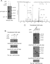
Tyr536 is a direct phosphorylation site for Lck but not for Csk. A, detection of Tyr(P)536 by phospho-mass spectrometry. Recombinant LYP-S227 was in vitro phosphorylated with Lck, and the resulting protein mixture was separated by SDS-PAGE and stained by Coomassie. The LYP protein band was excised and digested in gel by trypsin. The resulting peptide mixture was analyzed by nanoLC-MS/MS. The panel shows the MS/MS spectrum of the peptide spanning residues 517–548 of LYP, showing fragment ions that unambiguously pinpoint phosphorylation of residue Tyr536. B, Tyr536 is a major Lck phosphorylation site. Anti-HA IPs were performed from lysates of COS cells transfected with LYP-WT and LYP-F536 alone (lanes 1 and 2) or with Lck (lanes 3 and 4). C, Tyr536 is an unlikely Csk phosphorylation site. Anti-FLAG IPs were performed from lysates of COS cells transfected with Csk together with FLAG-tagged full-length LYP-S227 (lane 1) or the truncation mutants Δ288LYP (lane 2), Δ399LYP (lane 3), and Δ517LYP (lane 4).
FIGURE 8.
FIGURE 8.
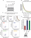
Reduced phosphorylation of Tyr536 on LYP-W620 leads to gain-of-function inhibition of TCR signaling. A, Tyr536 is more phosphorylated in LYP-R620 than LYP-W620. Anti-HA IPs were performed from lysates of JTAg cells transfected with LYP-R620 (lanes 1 and 2), LYP-W620 (lanes 3 and 4), LYP-R620/F536 (lanes 5 and 6), or LYP-W620/F536 (lanes 7 and 8) constructs. The cells were left unstimulated (lanes 1, 3, 5, and 7) or stimulated with C305 for 2 min (lanes 2, 4, 6, and 8). B–D, phosphorylation on Tyr536 inhibits LYP in TCR signaling. B, activation of an NFAT/AP1 reporter. JTAg cells were co-transfected with a 3xNFAT/AP1 firefly luciferase reporter, a control Renilla luciferase reporter, and LYP-R620, LYP-W620, LYP-R620/F536, or LYP-W620/F536. The cells were stimulated for 7 h with OKT3 and then lysed, and luciferase activity was measured on lysates. The average ± S.D. stimulation-induced increase in the ratio between firefly and Renilla luciferase activities of lysates of cells transfected with LYP-R620 (red squares and line), LYP-W620 (blue triangles and line), LYP-R620/F536 (green circles and line), or LYP-W620/F536 (yellow squares and line) was plotted versus LYP expression in same lysates as assessed by anti-HA blot. The lines are nonlinear fitting of data to an exponential decay equation, and 90% confidence intervals are shown (dashed lines). The data are representative of two experiments with similar results. C, induction of CD69. JTAg cells were transfected with HA-LYP-R620, HA-LYP-W620, HA-LYP-R620/F536, HA-LYP-W620/F536, HA-LYP-R620/S227, or vector alone. The cells were left unstimulated or stimulated with OKT3 for 4 h and were co-stained with an AlexaFluor488-conjugated anti-HA antibody and an allophycocyanin-conjugated anti-CD69 antibody. Live cells were gated by forward and side scatter and further gated for CD69 expression by comparison with the lower half of activated cells transfected with catalytically inactive LYP (HA-LYP-R620/S227). The corresponding percentage of gated T cells is shown in each box. Levels of overexpression of LYP mutants are shown as histograms of AlexaFluor488 fluorescence of HA-positive cells transfected with HA-LYP-R620 (red), HA-LYP-W620 (blue), HA-LYP-R620/F536 (green), HA-LYP-W620/F536 (yellow), or HA-LYP-R620/S227 (black). The gray-shaded graph shows HA-negative cells. D, phosphorylation on Tyr536 inhibits the phosphatase activity of LYP. JTAg cells were transfected with HA-LYP-R620, HA-LYP-W620, or HA-LYP-R620/F536. Anti-HA IPs were performed from lysates of cells stimulated with C305 for 2 min to maximize phosphorylation of LYP, and phosphatase activity was assessed continuously using the 14LckpCAP394 peptide as substrate. The histogram shows the average activity ± S.D. of IPed LYP-R620 (red column), LYP-W620 (blue column), or LYP-R620/F536 (green column) normalized for LYP expression by densitometric scanning of anti-HA blots of fractions of the IPs (see bottom panel). The time of reaction was optimized to ensure initial rate conditions and avoid any significant auto-dephosphorylation of the phosphatase (data not shown). The data are representative of two experiments with identical results.
FIGURE 9.
FIGURE 9.
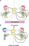
Model of regulation of LYP and Lck activity by phosphorylation of LYP on Tyr536.

Similar articles

Cited by

References

    1. Bottini N., Musumeci L., Alonso A., Rahmouni S., Nika K., Rostamkhani M., MacMurray J., Meloni G. F., Lucarelli P., Pellecchia M., Eisenbarth G. S., Comings D., Mustelin T. (2004) Nat. Genet. 36, 337–338 - PubMed
    1. Begovich A. B., Carlton V. E., Honigberg L. A., Schrodi S. J., Chokkalingam A. P., Alexander H. C., Ardlie K. G., Huang Q., Smith A. M., Spoerke J. M., Conn M. T., Chang M., Chang S. Y., Saiki R. K., Catanese J. J., Leong D. U., Garcia V. E., McAllister L. B., Jeffery D. A., Lee A. T., Batliwalla F., Remmers E., Criswell L. A., Seldin M. F., Kastner D. L., Amos C. I., Sninsky J. J., Gregersen P. K. (2004) Am. J. Hum. Genet. 75, 330–337 - PMC - PubMed
    1. Bottini N., Vang T., Cucca F., Mustelin T. (2006) Semin. Immunol. 18, 207–213 - PubMed
    1. Gregersen P. K., Lee H. S., Batliwalla F., Begovich A. B. (2006) Semin. Immunol. 18, 214–223 - PubMed
    1. Todd J. A., Walker N. M., Cooper J. D., Smyth D. J., Downes K., Plagnol V., Bailey R., Nejentsev S., Field S. F., Payne F., Lowe C. E., Szeszko J. S., Hafler J. P., Zeitels L., Yang J. H., Vella A., Nutland S., Stevens H. E., Schuilenburg H., Coleman G., Maisuria M., Meadows W., Smink L. J., Healy B., Burren O. S., Lam A. A., Ovington N. R., Allen J., Adlem E., Leung H. T., Wallace C., Howson J. M., Guja C., Ionescu-Tîrgovişte C., Simmonds M. J., Heward J. M., Gough S. C., Dunger D. B., Wicker L. S., Clayton D. G. (2007) Nat. Genet. 39, 857–864 - PMC - PubMed

Publication types

Substances

LinkOut - more resources