. 2010 Aug 6;107(3):398-407.
doi: 10.1161/CIRCRESAHA.110.222562. Epub 2010 Jun 10.

Alterations of L-type calcium current and cardiac function in CaMKII{delta} knockout mice

Affiliations

Alterations of L-type calcium current and cardiac function in CaMKII{delta} knockout mice

Lin Xu et al. Circ Res. .

Abstract

Rationale: Recent studies have highlighted important roles of CaMKII in regulating Ca(2+) handling and excitation-contraction coupling. However, the cardiac effect of chronic CaMKII inhibition has not been well understood.

Objective: We have tested the alterations of L-type calcium current (I(Ca)) and cardiac function in CaMKIIdelta knockout (KO) mouse left ventricle (LV).

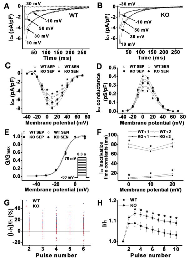
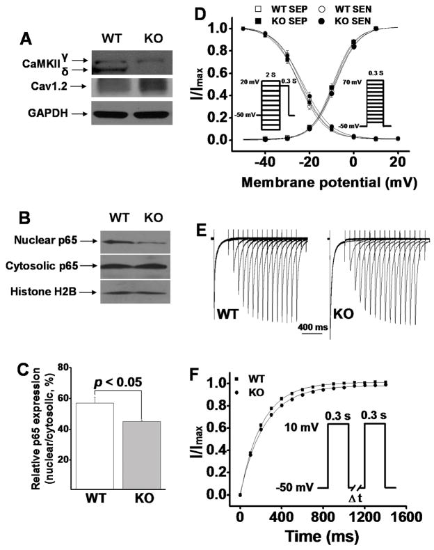
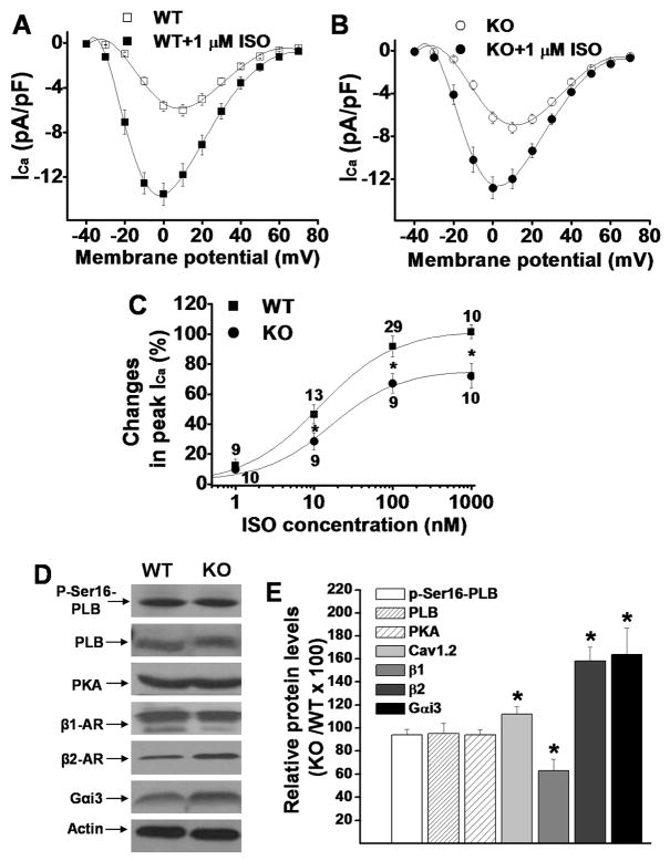
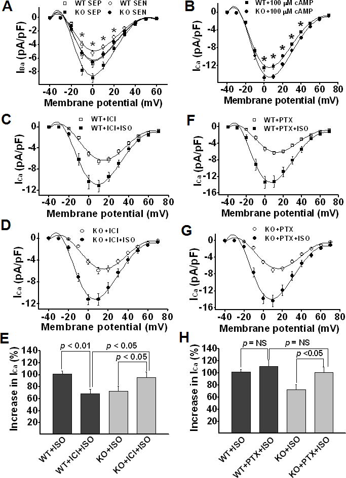
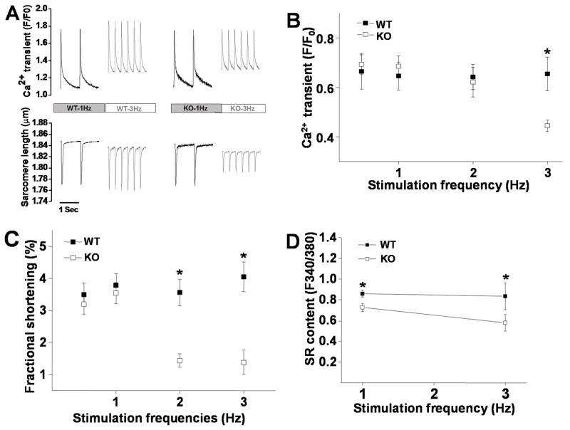
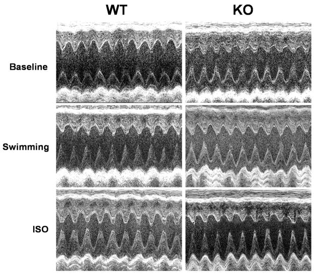
Methods and results: We used the patch-clamp method to record I(Ca) in ventricular myocytes and found that in KO LV, basal I(Ca) was significantly increased without changing the transmural gradient of I(Ca) distribution. Substitution of Ba(2+) for Ca(2+) showed similar increase in I(Ba). There was no change in the voltage dependence of I(Ca) activation and inactivation. I(Ca) recovery from inactivation, however, was significantly slowed. In KO LV, the Ca(2+)-dependent I(Ca) facilitation (CDF) and I(Ca) response to isoproterenol (ISO) were significantly reduced. However, ISO response was reversed by beta2-adrenergic receptor (AR) inhibition. Western blots showed a decrease in beta1-AR and an increase in Ca(v)1.2, beta2-AR, and Galphai3 protein levels. Ca(2+) transient and sarcomere shortening in KO myocytes were unchanged at 1-Hz but reduced at 3-Hz stimulation. Echocardiography in conscious mice revealed an increased basal contractility in KO mice. However, cardiac reserve to work load and beta-adrenergic stimulation was reduced. Surprisingly, KO mice showed a reduced heart rate in response to work load or beta-adrenergic stimulation.

Conclusions: Our results implicate physiological CaMKII activity in maintaining normal I(Ca), Ca(2+) handling, excitation-contraction coupling, and the in vivo heart function in response to cardiac stress.


Conflict of interest statement

Disclosures

There is no conflict to disclose.

Figures

Figure 1
Figure 1
A & B: Representative ICa traces recorded from WT and KO ventricular myocytes (SEN) by 300ms test pulses from a holding potential of −50mV to potentials between −30 to +50mV at pulse intervals of 10 s. ICa amplitude was increased and inactivation was accelerated at all test potentials in KO ventricular myocytes. C: ICa-voltage relationship in ventricular myocytes isolated from WT and CaMKIIδ KO LV. Current amplitudes were normalized to cell capacitance and plotted as mean values. D: Calcium channel conductance was normalized to cell capacitance and plotted as mean values. E: Normalized conductance-voltage relationship. F: Mean values of ICa inactivation time constants. τ1 and τ2 denote fast and slow inactivation time constants, respectively. G: Percentage changes of ICa at each stimulation pulse relative to the ICa at first pulse with 1 Hz stimulation. Blue and red dots represent changes of ICa from individual WT and KO myocytes, respectively. H: Normalized ICa facilitation from WT and the KO myocytes that showed positive ICa staircase. Vertical bars represent S.E.M.; * denotes p<0.05.
Figure 2
Figure 2
A: Representative Western blots showing ablated CaMKIIδ and increased Cav1.2 expression in CaMKIIδ KO LV. B: Western blots showing that NFkB component p65 nuclear translocation is reduced in KO LV. C: The mean values of relative nuclear portion p65 (nuclear/cytosolic, n=3) in WT and KO LV. D: Normalized voltage-dependence of ICa activation and inactivation. The recording protocols are shown as insets. E: Representative ICa recovery traces recorded (in SEN myocytes) from WT and KO ventricular myocytes, respectively. F: Mean values of normalized peak ICa (I/Imax) for ICa recovery from inactivation. The inset denotes recording protocol.
Figure 3
Figure 3
Panel A & B: Mean values of voltage-dependent ICa activation before and after ISO perfusion in ventricular myocytes isolated from WT and KO LV, respectively. Panel C: Dose-response relationship of peak ICa recorded in WT and KO LV in the presence of different concentrations of ISO. Panel D: Representative Western blot results for protein levels of p-Ser16-PLB, PLB, PKA, β1-AR, β2-AR, and Gαi3 (actin was used as loading control) in the WT and CaMKIIδ KO LV. The protein levels in KO LV relative to the levels in WT LV are statistically summarized in panel E (each value is a mean from 3 hearts). Vertical bars represent S.E.M. * denotes p<0.05, compared to proteins in WT.
Figure 4
Figure 4
Panel A: Mean values of IBa-voltage relationship. Panel B: Mean values of ICa recorded in WT and KO ventricular myocytes after internal diffusion of 100 μM 8-bromo-cAMP. Panel C&D: ISO effect on ICa recorded in WT and KO ventricular myocytes in the presence of β2-AR specific antagonist ICI118,551. Panel E: ISO (1 μM)-induced percent increase in peak ICa in WT and KO ventricular myocytes in the presence of ICI118,551. Panel F&G: ISO effect on ICa recorded in WT and KO ventricular myocytes in the presence of Gi inhibitor PTX. Panel H: ISO-induced percent increase in peak ICa in WT and KO ventricular myocytes in the presence of PTX. Vertical bars represent S.E.M.; * denotes p<0.05.
Figure 5
Figure 5
Panel A: Representative traces of Ca2+ transient and sarcomere shortening recorded at 1 and 3 Hz pacing rates in myocytes isolated from WT and KO LV. Panel B: Mean values of Ca2+ transient recorded in WT (n=15) and KO (n=13) ventricular myocytes at stimulation frequency of 0.5–3 Hz. Panel C: Mean values of sarcomere fractional shortening recorded in WT (n=15) and KO (n=13) ventricular myocytes at stimulation frequency of 0.5–3 Hz. Panel D: Mean values of SR Ca2+ contents recorded in WT (n=7) and KO (n=7) ventricular myocytes at stimulation frequency of 1 Hz and 3 Hz. Vertical bars represent S.E.M. * denotes p<0.05, compared to KO myocytes.
Figure 6
Figure 6
Representative images of short-axis two-dimensional M-mode echocardiography recorded from conscious WT and CaMKIIδ KO mice at baseline and after 15 minutes swimming or at 10 min after ISO injection, respectively.

Similar articles

Cited by

References

    1. Tobimatsu T, Fujisawa H. Tissue-specific expression of four types of rat calmodulin-dependent protein kinase II mRNAs. J Biol Chem. 1989;264:17907–17912. - PubMed
    1. Baltas LG, Karczewski P, Krause EG. The cardiac sarcoplasmic reticulum phospholamban kinase is a distinct delta-CaM kinase isozyme. FEBS Lett. 1995;373:71–75. - PubMed
    1. Singer HA, Benscoter HA, Schworer CM. Novel Ca2+/calmodulin-dependent protein kinase II gamma-subunit variants expressed in vascular smooth muscle, brain, and cardiomyocytes. J Biol Chem. 1997;272:9393–9400. - PubMed
    1. Hoch B, Haase H, Schulze W, Hagemann D, Morano I, Krause EG, Karczewski P. Differentiation-dependent expression of cardiac delta-CaMKII isoforms. J Cell Biochem. 1998;68:259–268. - PubMed
    1. Anderson ME. Calmodulin kinase and L-type calcium channels; a recipe for arrhythmias? Trends Cardiovasc Med. 2004;14:152–161. - PubMed

Publication types

MeSH terms

Substances