

An adjuvant for the induction of potent, protective humoral responses to an H5N1 influenza virus vaccine with antigen-sparing effect in mice
- PMID: 20538850
- PMCID: PMC2919013
- DOI: 10.1128/JVI.00596-10
An adjuvant for the induction of potent, protective humoral responses to an H5N1 influenza virus vaccine with antigen-sparing effect in mice
Abstract
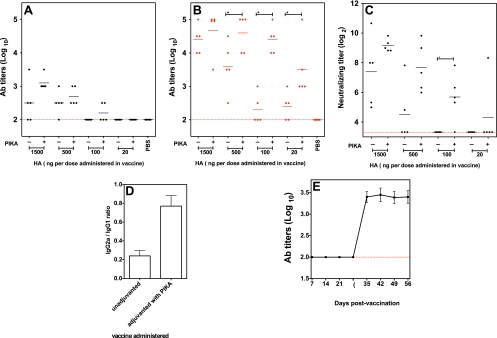
Intramuscular administration of inactivated influenza virus vaccine is the main vaccine platform used for the prevention of seasonal influenza virus infection. In clinical trials, inactivated H5N1 vaccines have been shown to be safe and capable of eliciting immune correlates of protection. However, the H5N1 vaccines are poorly immunogenic compared to seasonal influenza virus vaccines. Needle-free vaccination would be more efficient and economical in a pandemic, and the development of an effective and safe mucosal adjuvant will be an important milestone. A stabilized chemical analog of double-stranded RNA, PIKA, was previously reported to be a potent mucosal adjuvant in a murine model. While PIKA stimulates dendritic cells in vitro, little was known about its receptor and adjuvanting mechanism in vivo. In this study, we demonstrated that the immunostimulatory effect of PIKA resulted in an increased number of mature antigen-presenting cells, with the induction of proinflammatory cytokines at the inoculation site. In addition, coadministration of PIKA with a poorly immunogenic H5N1 subunit vaccine led to antigen sparing and quantitative and qualitative improvements of the immune responses over those achieved with an unadjuvanted vaccine in mice. The adjuvanted vaccine provided protection against lethal challenge with homologous and heterologous H5N1 wild-type viruses. Mice lacking functional TLR3 showed diminished cytokine production with PIKA stimulation, diminished antibody responses, and reduced protective efficacy against wild-type virus challenge following vaccination. These data suggest that TLR3 is important for the optimal performance of PIKA as an adjuvant. With its good safety profile and antigen-sparing effect, PIKA could be an attractive adjuvant for use in future pandemics.
Figures


Similar articles
-
Matrix-M adjuvanted virosomal H5N1 vaccine confers protection against lethal viral challenge in a murine model.Influenza Other Respir Viruses. 2011 Nov;5(6):426-37. doi: 10.1111/j.1750-2659.2011.00256.x. Epub 2011 May 9. Influenza Other Respir Viruses. 2011. PMID: 21668670 Free PMC article.
-
Broadly protective immunity against divergent influenza viruses by oral co-administration of Lactococcus lactis expressing nucleoprotein adjuvanted with cholera toxin B subunit in mice.Microb Cell Fact. 2015 Aug 5;14:111. doi: 10.1186/s12934-015-0287-4. Microb Cell Fact. 2015. PMID: 26242406 Free PMC article.
-
A study of Chitosan and c-di-GMP as mucosal adjuvants for intranasal influenza H5N1 vaccine.Influenza Other Respir Viruses. 2013 Nov;7(6):1181-93. doi: 10.1111/irv.12056. Epub 2012 Nov 21. Influenza Other Respir Viruses. 2013. PMID: 23170900 Free PMC article.
-
Prepandemic influenza vaccine H5N1 (split virion, inactivated, adjuvanted) [Prepandrix]: a review of its use as an active immunization against influenza A subtype H5N1 virus.BioDrugs. 2008;22(5):279-92. doi: 10.2165/00063030-200822050-00001. BioDrugs. 2008. PMID: 18778110 Review.
-
H5N1 vaccines in humans.Virus Res. 2013 Dec 5;178(1):78-98. doi: 10.1016/j.virusres.2013.05.006. Epub 2013 May 28. Virus Res. 2013. PMID: 23726847 Free PMC article. Review.
Cited by
-
TLR3-specific double-stranded RNA oligonucleotide adjuvants induce dendritic cell cross-presentation, CTL responses, and antiviral protection.J Immunol. 2011 Feb 15;186(4):2422-9. doi: 10.4049/jimmunol.1002845. Epub 2011 Jan 17. J Immunol. 2011. PMID: 21242525 Free PMC article.
-
Potent Neutralization Antibodies Induced by a Recombinant Trimeric Spike Protein Vaccine Candidate Containing PIKA Adjuvant for COVID-19.Vaccines (Basel). 2021 Mar 22;9(3):296. doi: 10.3390/vaccines9030296. Vaccines (Basel). 2021. PMID: 33810026 Free PMC article.
-
Advances in Adjuvanted Influenza Vaccines.Vaccines (Basel). 2023 Aug 21;11(8):1391. doi: 10.3390/vaccines11081391. Vaccines (Basel). 2023. PMID: 37631959 Free PMC article. Review.
-
Intratumoural immunotherapy: activation of nucleic acid sensing pattern recognition receptors.Immunooncol Technol. 2019 Oct 16;3:15-23. doi: 10.1016/j.iotech.2019.10.001. eCollection 2019 Oct. Immunooncol Technol. 2019. PMID: 35757301 Free PMC article. Review.
-
Antigen-activated dendritic cells ameliorate influenza A infections.J Clin Invest. 2013 Jul;123(7):2850-61. doi: 10.1172/JCI67550. Epub 2013 Jun 24. J Clin Invest. 2013. PMID: 23934125 Free PMC article.
References
-
- Beigel, J. H., J. Farrar, A. M. Han, F. G. Hayden, R. Hyer, M. D. de Jong, S. Lochindarat, T. K. Nguyen, T. H. Nguyen, T. H. Tran, A. Nicoll, S. Touch, and K. Y. Yuen. 2005. Avian influenza A (H5N1) infection in humans. N. Engl. J. Med. 353:1374-1385. - PubMed
-
- Bresson, J. L., C. Perronne, O. Launay, C. Gerdil, M. Saville, J. Wood, K. Hoschler, and M. C. Zambon. 2006. Safety and immunogenicity of an inactivated split-virion influenza A/Vietnam/1194/2004 (H5N1) vaccine: phase I randomised trial. Lancet 367:1657-1664. - PubMed
-
- Centers for Disease Control and Prevention. 2009. Swine influenza A (H1N1) infection in two children-Southern California, March-April 2009. MMWR Morb. Mortal. Wkly. Rep. 58:400-402. - PubMed
-
- Cleret, A., A. Quesnel-Hellmann, J. Mathieu, D. Vidal, and J. N. Tournier. 2006. Resident CD11c+ lung cells are impaired by anthrax toxins after spore infection. J. Infect. Dis. 194:86-94. - PubMed
Publication types
MeSH terms
Substances
Grants and funding
LinkOut - more resources
Full Text Sources
Other Literature Sources
Medical
