. 2010 Sep;84(17):8721-31.
doi: 10.1128/JVI.00144-10. Epub 2010 Jun 10.

Virus-induced Ca2+ influx extends survival of west nile virus-infected cells

Affiliations

Virus-induced Ca2+ influx extends survival of west nile virus-infected cells

Svetlana V Scherbik et al. J Virol. 2010 Sep.

Abstract

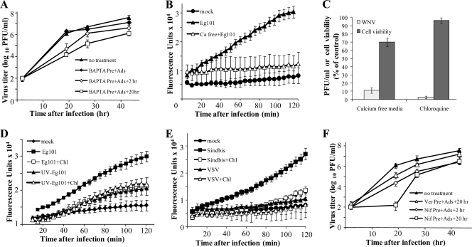
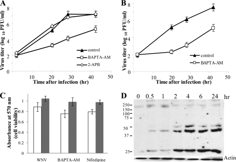
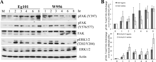
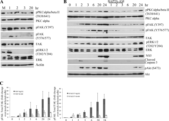
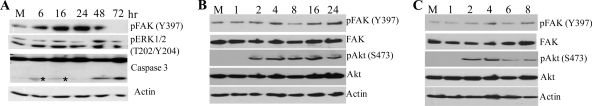
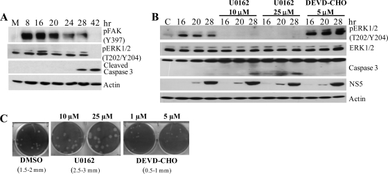
West Nile virus (WNV) infection leads to rapid and sustained Ca(2+) influx. This influx was observed with different strains of WNV and in different types of cells. Entry during virion endocytosis as well as through calcium channels contributed to the Ca(2+) influx observed in WNV-infected cells. Ca(2+) influx was not detected after infection with vesicular stomatitis virus (VSV) and occurred only through endocytosis in Sindbis virus-infected cells. Caspase 3 cleavage and activation of several kinases, including focal adhesion kinase (FAK), mitogen-activated extracellular signal-regulated protein kinase (ERK1/2), and protein-serine kinase B alpha (Akt), at early times after WNV infection were shown to be dependent on Ca(2+) influx. Although the activation of these kinases was sustained in virus-infected cells throughout infection, UV-inactivated WNV induced only a transient activation of FAK and ERK1/2 at early times after infection. The Ca(2+)-dependent FAK activation observed in WNV-infected cells was not mediated by alphavbeta3 integrins. Reduction of Ca(2+) influx at early times of infection by various treatments decreased the viral yield and delayed both the early transient caspase 3 cleavage and the activation of FAK, Akt, and ERK signaling. The results indicate that Ca(2+) influx is required for early infection events needed for efficient viral replication, possibly for virus-induced rearrangement of the endoplasmic reticulum (ER) membrane. Increased caspase 3 cleavage at both early (transient) and late times of infection correlated with decreased activation of the FAK and ERK1/2 pathways, indicating a role for these kinases in extending the survival of flavivirus-infected cells.


Figures

FIG. 1.
FIG. 1.
WNV infection induces an extracellular Ca2+ influx that enhances virus production. Confluent monolayers of C3H/He MEFs were pretreated with 50 μM BAPTA (A), preincubated in a calcium-free medium (B), or treated with 20 μM chloroquine (D) or 50 μM nifedipine (F) for 2 h. The cells were then mock infected or infected with WNV at an MOI of 5 for 1 h in the presence of drug and were maintained thereafter in medium with drug (or calcium-free medium) for either 2 or 20 h after infection, followed by replacement with fresh medium. Samples of culture fluid were harvested at the indicated times, and titers were determined by plaque assay on BHK cells. Error bars indicate ± standard deviations (SD) (n = 3). (C) The viability of chloroquine-treated MEFs or cells maintained in calcium-free medium was determined by MTT assay as described in Materials and Methods. Extracellular virus titers 24 h after infection were determined by plaque assay on BHK cells. Data are shown as percentages of control values. (B, D, and E) C3H/He MEFs in 96-well plates were preloaded with Fluo-4NW (Molecular Probes) for 30 min at 37°C. The cells were then either mock infected, infected with WNV Eg101 at an MOI of 5 (B and D) or with Sindbis virus or VSV (MOI of 5) (E), or infected in the presence of 20 μM chloroquine. (D) Cells were incubated with UV-inactivated WNV Eg101 (equivalent to an MOI of 5) in the presence or absence of chloroquine (Chl). Fluorescence was measured at 5-min intervals in a Victor 3 1420 multilabel plate reader (Perkin Elmer). The data points are averages for six measurements.
FIG. 2.
FIG. 2.
Intracellular Ca2+ is important for efficient WNV production. Confluent monolayers of C3H/He (A) or C57BL/6 (B) MEFs were pretreated with 25 μM BAPTA-AM or 100 μM 2-APB (A) for 0.5 h. The cells were then either mock infected or infected with WNV at an MOI of 5 for 1 h in the presence of drug and were maintained thereafter in medium with drug for 2 h after infection, followed by replacement with fresh medium. Samples of culture fluid were harvested at the indicated times, and titers were determined by plaque assay on BHK cells. Error bars indicate ±SD (n = 3). Control, 0.1% DMSO. (C) Cell viability was measured by MTT assay. White bars, C3H/He MEFs; gray bars, BHK cells infected with WNV Eg101 and treated with BAPTA-AM (as described in the legend to Fig. 1) or nifedipine (20 h). (D) C3H/He MEFs were serum starved for 3 h before infection with WNV Eg101 at an MOI of 10. Cell extracts were prepared 30 min and 1, 2, 4, and 6 h after infection and were analyzed by immunoblotting with an anti-P-Tyr antibody.
FIG. 3.
FIG. 3.
WNV infection induces phosphorylation of FAK and ERK1/2. (A) BHK cells were infected with WNV Eg101 or W956 at an MOI of 5. At the indicated times after infection, cell lysates were prepared and proteins were separated by 10% SDS-PAGE, transferred to membranes, and analyzed by Western blotting, using antibodies specific for the indicated protein phosphorylation sites, as described in Materials and Methods. Actin was used as a loading control. (B) Results obtained from three independent experiments are summarized graphically as fold changes in the ratio of optical density units (ODU) of phosphorylated FAK to ODU of total FAK divided by the ratio for mock-infected cells. Error bars indicate ±SD.
FIG. 4.
FIG. 4.
WNV infection induces FAK and ERK1/2 phosphorylation and increases intracellular Ca2+ levels in CS-1 hamster melanoma cells. (A) CS-1 cells infected with WNV Eg101 at an MOI of 5. Samples of culture fluid were harvested at the indicated times, and titers were determined by plaque assay on BHK cells. Error bars indicate ±SD (n = 3). (B) CS-1 cells seeded in 96-well plates were loaded with Fluo-4NW for 30 min at 37°C. The cells were then incubated with WNV Eg101 at an MOI of 5 or mock infected for 1 h. Fluorescence was measured at 5-min intervals in a Victor 3 1420 multilabel plate reader (Perkin Elmer). The averages of three measurements were plotted. (C) CS-1 cells infected with WNV Eg101 at an MOI of 5. Cell lysates were harvested at the indicated times after infection. Proteins were separated by SDS-PAGE and analyzed by Western blotting, using antibodies specific for the indicated protein sites, as described in Materials and Methods. The total ERK proteins were used as the loading control.
FIG. 5.
FIG. 5.
Sustained WNV-mediated activation of FAK requires replicating virus and is Ca2+ dependent. C3H/He MEFs were incubated with UV-inactivated WNV Eg101 (equivalent to an MOI of 5) (A) or were mock infected, infected with WNV at an MOI of 5 for the indicated periods, or infected in the presence of 25 μM BAPTA-AM and maintained thereafter in medium with drug for 2 h after infection, followed by replacement with fresh medium (B and C). Cell lysates were prepared at the indicated times after infection. Proteins were separated by SDS-PAGE and analyzed by Western blotting, using antibodies specific for the indicated protein sites. Actin was used as a loading control. (C) Results obtained from three independent experiments are summarized graphically as fold changes in the ratio of ODU of phosphorylated FAK to ODU of total FAK divided by the ratio for mock-infected cells. Error bars indicate ±SD.
FIG. 6.
FIG. 6.
WNV infections, but not VSV or Sindbis virus infections, induce phosphorylation of FAK. C3H/He MEFs were mock infected or infected with WNV at an MOI of 5 for the indicated periods (A) or were infected with Sindbis virus (B) or VSV (C) at an MOI of 10. Cell lysates were prepared at the indicated times after infection. Proteins were separated by SDS-PAGE and analyzed by Western blotting, using antibodies specific for the indicated protein sites. Asterisks indicate nonspecific bands. Actin was used as a loading control.
FIG. 7.
FIG. 7.
Detection of ERK1/2 activation and caspase 3 cleavage. (A) BHK cells were infected with WNV Eg101 at an MOI of 5 and then incubated with medium containing either 0.1% DMSO (solvent), 10 or 25 μM U0126, or 5 μM DEVD-CHO. At the indicated times after infection, cell lysates were prepared. Proteins were separated by SDS-PAGE and analyzed by Western blotting, using antibodies specific for the indicated protein phosphorylation sites. Actin was used as a loading control. The blots shown are representative of the results obtained from three independent experiments. (B) BHK cells were infected with WNV Eg101 at an MOI of 5. Cell lysates were harvested at the indicated times after infection. Proteins were separated by SDS-PAGE and analyzed by Western blotting, using antibodies specific for the indicated protein sites, as described in Materials and Methods. Actin was used as a loading control. (C) Effects of incubation of infected cells with MEK and caspase 3 inhibitors on plaque size. Confluent monolayers of BHK cells were infected with serial 10-fold dilutions of WNV and incubated under 1% agarose for 72 h in the presence of DMSO or one of the indicated inhibitors. The plaque size is shown for each condition.

References

    1. Achison, M., C. M. Elton, P. G. Hargreaves, C. G. Knight, M. J. Barnes, and R. W. Farndale. 2001. Integrin-independent tyrosine phosphorylation of p125(fak) in human platelets stimulated by collagen. J. Biol. Chem. 276:3167-3174. - PubMed
    1. Alessandro, R., L. Masiero, K. Lapidos, J. Spoonster, and E. C. Kohn. 1998. Endothelial cell spreading on type IV collagen and spreading-induced FAK phosphorylation is regulated by Ca2+ influx. Biochem. Biophys. Res. Commun. 248:635-640. - PubMed
    1. Alfano, M., H. Schmidtmayerova, C. A. Amella, T. Pushkarsky, and M. Bukrinsky. 1999. The B-oligomer of pertussis toxin deactivates CC chemokine receptor 5 and blocks entry of M-tropic HIV-1 strains. J. Exp. Med. 190:597-605. - PMC - PubMed
    1. Beasley, D. W., L. Li, M. T. Suderman, and A. D. Barrett. 2002. Mouse neuroinvasive phenotype of West Nile virus strains varies depending upon virus genotype. Virology 296:17-23. - PubMed
    1. Berridge, M. J., M. D. Bootman, and H. L. Roderick. 2003. Calcium signalling: dynamics, homeostasis and remodelling. Nat. Rev. Mol. Cell. Biol. 4:517-529. - PubMed

Publication types