. 2010 Sep;84(17):8712-20.
doi: 10.1128/JVI.00523-10. Epub 2010 Jun 10.

Hit-and-run stimulation: a novel concept to reactivate latent HIV-1 infection without cytokine gene induction

Affiliations

Hit-and-run stimulation: a novel concept to reactivate latent HIV-1 infection without cytokine gene induction

Frank Wolschendorf et al. J Virol. 2010 Sep.

Abstract

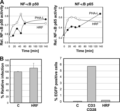
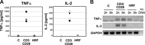
Current antiretroviral therapy (ART) efficiently controls HIV-1 replication but fails to eradicate the virus. Even after years of successful ART, HIV-1 can conceal itself in a latent state in long-lived CD4(+) memory T cells. From this latent reservoir, HIV-1 rebounds during treatment interruptions. Attempts to therapeutically eradicate this viral reservoir have yielded disappointing results. A major problem with previously utilized activating agents is that at the concentrations required for efficient HIV-1 reactivation, these stimuli trigger high-level cytokine gene expression (hypercytokinemia). Therapeutically relevant HIV-1-reactivating agents will have to trigger HIV-1 reactivation without the induction of cytokine expression. We present here a proof-of-principle study showing that this is a possibility. In a high-throughput screening effort, we identified an HIV-1-reactivating protein factor (HRF) secreted by the nonpathogenic bacterium Massilia timonae. In primary T cells and T-cell lines, HRF triggered a high but nonsustained peak of nuclear factor kappa B (NF-kappaB) activity. While this short NF-kappaB peak potently reactivated latent HIV-1 infection, it failed to induce gene expression of several proinflammatory NF-kappaB-dependent cellular genes, such as those for tumor necrosis factor alpha (TNF-alpha), interleukin-8 (IL-8), and gamma interferon (IFN-gamma). Dissociation of cellular and viral gene induction was achievable, as minimum amounts of Tat protein, synthesized following application of a short NF-kappaB pulse, triggered HIV-1 transactivation and subsequent self-perpetuated HIV-1 expression. In the absence of such a positive feedback mechanism, cellular gene expression was not sustained, suggesting that strategies modulating the NF-kappaB activity profile could be used to selectively trigger HIV-1 reactivation.


Figures

FIG. 1.
FIG. 1.
Culture supernatants from Massilia timonae reactivate latent HIV-1 infection. Latently HIV-1-infected CA5 reporter T cells were treated with 10 μl of Massilia timonae cell culture supernatant (HRF). Untreated control cells and HRF-treated cells were subjected to flow cytometric analysis. (A and C) FSC/SSC dot plots were used to determine cell viability as a function of changes in the FSC (cell size) and SSC (granularity) phenotypes of the cells. (B and D) EGFP expression was used as a direct and quantitative marker of HIV-1 expression at 24 h poststimulation. (E) To demonstrate the reproducibility of the observed effect, we tested three independent preparations of HRF (M. timonae #1 to #3) on four different latently infected T-cell lines (5F3, 8E12, CA5, and 12D4). The percentages of EGFP-positive cells following stimulation are depicted as histograms. Cell culture supernatants of Pseudomonas aeruginosa cultures grown under similar conditions were used as a specificity control. (F) CA5 cells were stimulated with either PMA, TNF-α, or HRF, and HIV-1 Gag p24 levels were determined by ELISA to demonstrate viral protein production following HRF stimulation of CA5 T cells. C, control. (G) HRF-mediated reactivation is not the result of pyrogenic activity. Since Massilia timonae is a Gram-negative bacterium, we tested whether HIV-1 reactivation could be triggered by endotoxin-like activities. To remove endotoxins from the HRF preparations, we incubated the Massilia timonae culture supernatants with polymyxin B-agarose prior to stimulation of the latently HIV-1-infected CA5 reporter T cells with increasing doses of the HRF preparations. Levels of HIV-1 reactivation were determined as the percentage of EGFP-positive cells by flow cytometric analysis at 48 h poststimulation. (H) Latently HIV-1-infected CA5 reporter T cells were treated with increasing concentrations of HRF or LPS. The functionality of LPS was demonstrated by stimulating latently HIV-1-infected monocytic THP89GFP cells with increasing amounts of LPS and then measuring the level of LPS-mediated HIV-1 reactivation. For all conditions, levels of HIV-1 reactivation were determined as the percentage of EGFP-positive cells at 48 h poststimulation, using flow cytometric analysis.
FIG. 2.
FIG. 2.
HIV-1 reactivation in CA5 T cells triggered by coculture with Massilia timonae. Mixtures of M. timonae or E. coli and a constant number of latently HIV-1-infected CA5 reporter T cells (1 × 10⁶ cells) were cocultured at different ratios for 24 h. (A, C, and E) Using a logarithmic representation of the FSC/SSC analysis, it was possible to visualize M. timonae, E. coli, and the T cells in the same FSC/SSC dot plot, using flow cytometric analysis. (B, D, and F) By gating on the T-cell population, we could determine the level of HIV-1 reactivation in latently HIV-1-infected CA5 T cells as a function of the number of bacteria. M. timonae cocultures with T cells were started with a very small (A and B) and a very large (C and D) number of bacteria, both of which provided full HIV-1 reactivation at the population level. (E and F) E. coli cocultures with CA5 T cells were started at the same low bacterium/T-cell ratio as that used for M. timonae cocultures in panel A, but they did not reactivate the latent HIV-1 virus. FSC/SSC plots were used to determine any changes in cell numbers, cell morphology, or viability. The surface of the E. coli dot plot demonstrates massive bacterial growth. EGFP fluorescence was determined as a direct marker of HIV-1 expression. RFP, secondary fluorescence.
FIG. 3.
FIG. 3.
HRF triggers suboptimal functional induction of NF-κB-dependent gene expression. (A) The HIV-1-reactivating kinetics of HRF relative to those of known HIV-1-reactivating agents that signal through the NF-κB pathway were determined by stimulating CA5 T cells with optimal concentrations of HRF, PMA, and TNF-α and analyzing HIV-1 reactivation as a function of EGFP expression over a period of 48 h. Reactivation data for all agents are depicted as percentages of EGFP-positive cells. (B) To test the ability of HRF to mediate Tat-independent activation of the HIV-1 LTR, NOMI cells were stimulated with increasing concentrations of PMA (0.01 to 10 ng/ml), TNF-α (0.003 to 30 ng/ml), and HRF supernatant (0.3 to 100 μl). The level of induction of the stably integrated HIV-1 LTR reporter construct was then measured as the EGFP mean channel fluorescence (MCF) detectable after 24 h, as determined by flow cytometric analysis. Arrows indicate the concentrations at which the respective stimuli would have triggered full HIV-1 reactivation in the latently HIV-1-infected CA5 T-cell line. (C) 293T cells were transfected with several NF-κB-dependent promoter constructs and then stimulated with either TNF-α (10 ng/ml) or HRF (25 μl). Promoter induction was measured as total EGFP expression. NF-κB, pNF-κB-d2EGFP; LTR, HIV-1 LTR-GFP; IL-8, pIL-8 GFP; TNF, pTNF-GFP; MSCV, murine stem cell virus LTR-driven GFP (no NF-κB-responsive elements).
FIG. 4.
FIG. 4.
Kinetics of HRF-mediated induction of NF-κB activity. Jurkat T cells or latently HIV-1-infected CA5 T cells were stimulated with optimal concentrations of PMA or HRF, and cells were harvested at the indicated time points. Nuclear extracts were generated, and NF-κB p50 activity (A and C) and NF-κB p65 activity (B and D) were determined for the indicated time points, using a TransAM NF-κB family ELISA kit. To demonstrate specificity and that active NF-κB complexes would also bind to the HIV-1 NF-κB element, we performed cold competition experiments, using an NF-κB consensus oligonucleotide (NF-κB), an oligonucleotide representing the HIV-1 NL43 NF-κB element (NL43), and a scrambled oligonucleotide sequence (Scr). Data for NF-κB p50 are depicted in panel E, and those for NF-κB p65 are depicted in panel F.
FIG. 5.
FIG. 5.
Altered kinetic IκB regulation following HRF stimulation. (A) Jurkat T cells were stimulated with either TNF-α or HRF and harvested at the indicated time points. Western blots were performed to determine the kinetics of changes in IκBα expression levels. (B) For the first 2 h, the time resolution was sufficiently high to exactly determine the oscillation of IκBα expression (solid lines). After this time point, changes in IκBα expression for both stimuli were fitted to the determined IκBα expression levels at the 4-h and 6-h time points by assuming similar oscillation amplitudes and frequencies for both stimuli (dotted lines). Anti-tubulin antibodies were used to confirm equal preparation qualities and proper loading.
FIG. 6.
FIG. 6.
Effects of HRF stimulation of primary T cells on NF-κB activation and susceptibility to HIV-1 infection. (A) PBMCs were activated with optimal concentrations of either PHA-L or HRF. Cells were harvested at the indicated time points. Nuclear extracts were generated, and NF-κB p50 activity and NF-κB p65 activity were determined using a TransAM NF-κB family ELISA kit. (B) PBMCs from four different healthy donors were stimulated with an anti-CD3/CD28 antibody combination and infected with an EGFP reporter virus on day 4 poststimulation. HIV-1 replication was monitored in the absence (control) or presence of HRF for 5 days, and the achieved HIV-1 infection levels were determined by flow cytometric analysis of the percentage of EGFP-positive cells. All infections were normalized to the infection level in the untreated culture, and the histogram presents the mean infection level obtained for 4 donor cultures ± the standard deviation (left panel). PBMCs isolated from healthy donors were left unstimulated (C), stimulated with an anti-CD3/CD28 MAb combination, or treated with HRF and infected with an EFGP reporter virus. HIV-1 infection levels were determined as the percentage of EGFP-positive cells on day 2 postinfection (right panel).
FIG. 7.
FIG. 7.
Effects of HRF stimulation on cytokine expression in PBMCs. (A) PBMCs from four different healthy donors were left unstimulated (C), stimulated with an anti-CD3/CD28 MAb combination as a positive control, or stimulated with a concentration of HRF that would trigger maximum HIV-1 reactivation in CA5 T cells. Culture supernatants were collected after 24 h, and the concentrations of a panel of cytokines were determined by BioPlex analysis. Concentrations of the proinflammatory cytokines TNF-α and IL-2 are presented for four individual donors. (B) To test whether the lack of cytokine production at the protein level was correlated with low-level mRNA transcription, PBMCs that were stimulated with either an anti-CD3/CD28 MAb combination or HRF were harvested at the indicated time points. mRNAs were isolated and used to generate cDNAs. Specific primer pairs for IL-2 and TNF-α were used to demonstrate the presence or absence of cytokine-specific mRNA at the indicated time points. Glyceraldehyde-3-phosphate dehydrogenase (GAPDH) primers were used to control for cDNA quality. The depicted PCRs are representative of three independent experiments using PBMCs from three different healthy donors.

Similar articles

Cited by

References

    1. Choi, C., O. Kutsch, J. Park, T. Zhou, D. W. Seol, and E. N. Benveniste. 2002. Tumor necrosis factor-related apoptosis-inducing ligand induces caspase-dependent interleukin-8 expression and apoptosis in human astroglioma cells. Mol. Cell. Biol. 22:724-736. - PMC - PubMed
    1. Chun, T. W., L. Carruth, D. Finzi, X. Shen, J. A. DiGiuseppe, H. Taylor, M. Hermankova, K. Chadwick, J. Margolick, T. C. Quinn, Y. H. Kuo, R. Brookmeyer, M. A. Zeiger, P. Barditch-Crovo, and R. F. Siliciano. 1997. Quantification of latent tissue reservoirs and total body viral load in HIV-1 infection. Nature 387:183-188. - PubMed
    1. Chun, T. W., R. T. Davey, Jr., M. Ostrowski, J. S. Justement, D. Engel, J. I. Mullins, and A. S. Fauci. 2000. Relationship between pre-existing viral reservoirs and the re-emergence of plasma viremia after discontinuation of highly active anti-retroviral therapy. Nat. Med. 6:757-761. - PubMed
    1. Chun, T. W., D. Engel, M. M. Berrey, T. Shea, L. Corey, and A. S. Fauci. 1998. Early establishment of a pool of latently infected, resting CD4(+) T cells during primary HIV-1 infection. Proc. Natl. Acad. Sci. U. S. A. 95:8869-8873. - PMC - PubMed
    1. Chun, T. W., D. Engel, S. B. Mizell, L. A. Ehler, and A. S. Fauci. 1998. Induction of HIV-1 replication in latently infected CD4+ T cells using a combination of cytokines. J. Exp. Med. 188:83-91. - PMC - PubMed

Publication types

MeSH terms