. 2010 Aug;84(16):8098-110.
doi: 10.1128/JVI.00966-10. Epub 2010 Jun 10.

Crystal structure of PG16 and chimeric dissection with somatically related PG9: structure-function analysis of two quaternary-specific antibodies that effectively neutralize HIV-1

Affiliations

Crystal structure of PG16 and chimeric dissection with somatically related PG9: structure-function analysis of two quaternary-specific antibodies that effectively neutralize HIV-1

Marie Pancera et al. J Virol. 2010 Aug.

Abstract

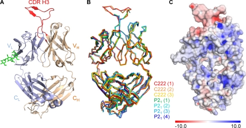
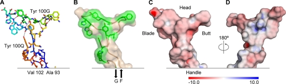
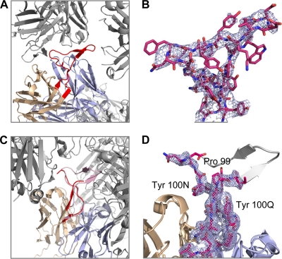
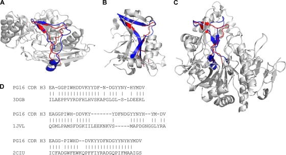
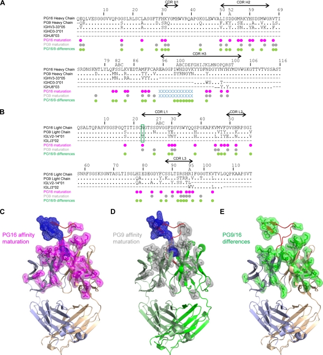
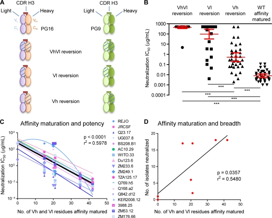
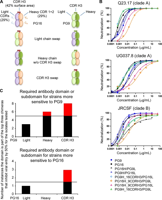
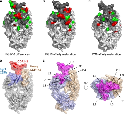
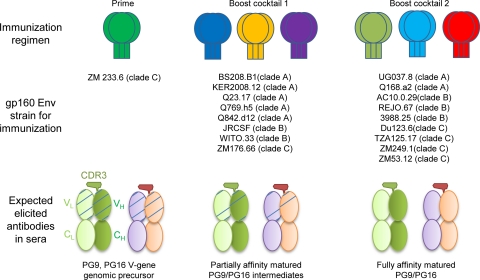
HIV-1 resists neutralization by most antibodies. Two somatically related human antibodies, PG9 and PG16, however, each neutralize 70 to 80% of circulating HIV-1 isolates. Here we present the structure of the antigen-binding fragment of PG16 in monoclinic and orthorhombic lattices at 2.4 and 4.0 A, respectively, and use a combination of structural analysis, paratope dissection, and neutralization assessment to determine the functional relevance of three unusual PG9/PG16 features: N-linked glycosylation, extensive affinity maturation, and a heavy chain-third complementarity-determining region (CDR H3) that is one of the longest observed in human antibodies. Glycosylation extended off the side of the light chain variable domain and was not required for neutralization. The CDR H3 formed an axe-shaped subdomain, which comprised 42% of the CDR surface, with the axe head looming approximately 20 A above the other combining loops. Comprehensive sets of chimeric swaps between PG9 and PG16 of light chain, heavy chain, and CDR H3 were employed to decipher structure-function relationships. Chimeric swaps generally complemented functionally, with differences in PG9/PG16 neutralization related primarily to residue differences in CDR H3. Meanwhile, chimeric reversions to genomic V genes showed isolate-dependent effects, with affinity maturation playing a significant role in augmenting neutralization breadth (P = 0.036) and potency (P < 0.0001). The structural and functional details of extraordinary CDR H3 and extensive affinity maturation provide insights into the neutralization mechanism of and the elicitation pathway for broadly neutralizing antibodies like PG9 and PG16.


Figures

FIG. 1.
FIG. 1.
Crystal structure of the antigen-binding fragment (Fab) of antibody PG16. Anti-HIV-1 antibodies that effectively neutralize HIV-1 often have unusual structural characteristics. With PG16, an extraordinary CDR H3 forms a separate subdomain, which towers over the combining-region surface. (A) Structure of antibody PG16, with complete combining region. A Cα-ribbon representation shows the light chain in blue, the heavy chain in tan, and its CDR H3 in red. The variable domains comprise the top half of the image, and the constant regions comprise the bottom. The single N-linked glycan (shown in green stick representation) was sufficiently well ordered at its protein-proximal base to define the placement of three sugars. The displayed Fab molecule is molecule 1 from the C222 lattice as shown in panel B. The CDR H3 of PG16 dominates the combining site and comprises 42% of the total surface area of the complementarity-determining regions. (B) Superposition of asymmetric unit components. PG16 crystallized in orthorhombic (C222) and monoclinic (P21) lattices, with 3 and 4 molecules per asymmetric unit, respectively. Shown are the Cα-backbone traces for all seven of the crystallized forms of antibody PG16, after superposition of variable domain-framework regions. The number for each molecule in each lattice is indicated in parentheses. (C) PG16 Fab electrostatic surface potentials. The PG16 electrostatic potentials are displayed on the molecular surface of PG16, with blue showing positively charged surfaces and red showing negatively charged surfaces. The scale is from −10 to +10 kT/e.
FIG. 2.
FIG. 2.
Structural and chemical properties of the PG16 CDR H3. One of the longest human CDR H3s ever observed, the PG16 CDR H3 is anionic and secured by a number of hydrogen bonds but lacks the hydrophobic core associated with more stable configurations. (A) Stick representation with rainbow coloring of carbon atoms from blue to red, Ala93 to Val102, respectively. Hydrogen bonds are depicted as dotted lines. Backbone hydrogen bonds include NGly98-OTyr100O, OGly98-NTyr100O, NIle100-OTyr100G, OIle100-NTyr100G, NTrp100A-OVal100E, OHis100B-NVal100E, NAsp100I-OAla100M, and OAsp100I-NAsp100L residue numbering follows the Kabat format (23). (B) Transparent molecular surface over a backbone ribbon with the side chains of aromatic residues shown as sticks and colored green. The CDR H3 extends from strand F and returns into strand G of the Fab. (C) Molecular surface colored according to electrostatic potentials from −10 to +10 kT/e, red to blue, respectively, with analogy to the axe vocabulary indicated. (D) 180° rotation of panel C about the y axis.
FIG. 3.
FIG. 3.
CDR H3 flexibility and tyrosine clasp. The head of the CDR H3 appears to have the ability to deform substantially in response to different environments, perhaps enabling it to maneuver into a recessed epitope on HIV-1. In contrast, the handle of the CDR H3 appears invariant, fixed by Tyr100N and Tyr100Q, which embrace and support the CDR H3 N terminus. (A) Ordered CDR H3 as seen in the C222 lattice. Neighboring molecules are colored gray whereas the CDR H3 is highlighted in red. (B) The CDR H3 of PG16 Fab from the C222 lattice is shown in stick representation, and the 2Fo-Fc map obtained after rigid body and TLS refinement in PHENIX is shown in light blue at 1σ. (C) Disordered CDR H3 (red) as seen in the P21 lattice. The ordered CDR H3 (pink) from the C222 lattice has been superimposed to highlight potential clashes within the crystal lattice. (D) A close-up of the CDR H3 (in red sticks) from the P21 lattice is shown to highlight the tyrosine clasp. The missing portion of the CDR H3 loop is modeled in gray. 2Fo-Fc electron density surrounding the CDR H3 loop is also shown in light blue at 1σ.
FIG. 4.
FIG. 4.
Structural homologs of PG16 CDR H3. Analysis of structural homologs allows insight into the function of the PG16 subdomain in other contexts. Six structural homologs were identified from a database of 16,938 proteins. Based on the structural similarity, they could be categorized into three families, and a single example of each family is shown. Regions of homology to PG16 are highlighted in blue, and the CDR H3 of PG16 is shown in red. (A) One family consisted of three proteins, two from the enolase superfamily and one with unknown function (PDB IDs 2NQL, 3DGB, and 2OZ8). 3DGB is depicted here. The region of structural homology to the CDR H3 was packed on the protein surface, and the RMSD of aligned residues ranged from 3.47 to 3.71 Å. (B) In a second family, the CDR H3 was matched to a buried region sandwiched by secondary structure elements on both sides in two pyrophosphorylase proteins (PDB IDs 1JV1 and 2YQC), with an RMSD of 2.16 to 2.26 Å. 1JV1 is shown as the example of the second family. (C) The third family had only one protein, 2ICU, in which the CDR H3 of PG16 matched to a core segment surrounded by other structural components with an RMSD of 3.0 Å. None of the structural homologs protrudes from the protein body as does the CDR H3 of PG16. (D) Sequence alignments of the PG16 CDR H3 with the three structural homologs shown in panels A to C. The residues that have been aligned between the CDR H3 of PG16 and the structural homolog to calculate RMSD are shown with vertical lines.
FIG. 5.
FIG. 5.
Affinity maturation and sequence differences between PG9 and PG16. Somatic hypermutation alters residues throughout the variable domains of both PG9 and PG16. (A) Sequence alignments of PG16 and PG9 heavy chain with genomic precursor gene (Vh, D, and Jh) using ClustalW2 (28). (B) Sequence alignments of PG16 and PG9 light chain with genomic precursor gene (Vl and Jl). Dots indicate identical residues, and a dash indicates that there is no corresponding sequence for that region. PG16 and PG9 affinity maturation from germ line is shown in magenta and gray dots, respectively. The blue crosses (“x”) indicate the residues that are not included in the Vh or D gene. The site of glycosylation is boxed in green on the light chain. PG9 and PG16 differences are shown with green dots. The sequences of the CDRs are indicated by lines with arrowheads. (C) Structural mapping of PG16 affinity maturation changes. Cα-ribbon representation of PG16 Fab with somatic mutations shown in magenta (stick and transparent surface representation). The CDR H3 residues not included in the Vh or D alignment are shown in blue mesh. The CDR H3 is shown in red, the PG16 light chain in light blue, and the PG16 heavy chain in tan. (D) Structural mapping of PG9 affinity maturation changes. Cα-ribbon representation of PG9 Fab homology model with somatic mutations shown in gray (stick and transparent surface representation). The CDR H3 residues not included in the Vh or Dh alignment are shown in blue mesh. The CDR H3 is shown in red, the PG9 light chain in light green, and the PG9 heavy chain in green. (E) Structural mapping of PG9 and PG16 sequence differences. Cα-ribbon representation of the PG16 Fab structure with CDR H3, light chain, and heavy chain colored as in panel C. Residues where PG9 differs from PG16 are displayed in green stick and transparent surface representation.
FIG. 6.
FIG. 6.
V-gene reverted chimeras and dissection of functional relevance. Resolution-enhancing methods are particularly useful when analyzing diffuse effects such as somatic hypermutation, which alters ∼50 residues between PG9 and PG16. (A) Schematic V-gene reverted chimeras of PG16 and PG9 Fabs are shown: both heavy and light chain V genes reverted to germ line precursor, only heavy V gene reverted, or only light chain V gene reverted (see Fig. S2 in the supplemental material). PG16 light chain is in light blue, PG16 heavy chain is in tan, and PG16 CDR H3 is in red; PG9 light chain is in light green, PG9 heavy chain is in green, and PG9 CDR H3 is bordered in dark green. V-gene reversion to germ line is shown by hatching. (B) IC50 are reported for each category of antibodies: V-gene heavy and light chain reverted to germ line (VhVl), Vl reverted, Vh reverted, and affinity-matured antibodies, i.e., wild-type PG9 and PG16. There were significant differences in IC50 among the four groups according to a Kruskal-Wallis (KW) test (KW = 112, P < 0.0001) or a one-way analysis of variance (F = 129.3, P < 0.0001). Dunn's multiple comparison post hoc tests indicated that differences between the groups were highly significant (P < 0.001, indicated by a triple asterisk on the graph). (C) Correlation between affinity maturation and potency. The IC50 (potency) values were plotted as a function of the number of residues that are affinity matured, i.e., 15 and 21 residues for the Vl of PG9 and PG16, respectively, and 20 residues for the Vh of both PG9 and PG16. The Vh and Vl reverted chimeras contain no affinity maturation in the V gene, and the PG9 and PG16 have 35 and 41 residues affinity matured in the V gene, respectively. Analysis indicated that one isolate, REJO, could not be fitted to a linear regression (see Materials and Methods). The rest of the data could be fitted by linear regression, and statistical analysis using Prism (version 5.00 for Windows; GraphPad) indicated that it was valid to model the entire data set (minus REJO) with a single linear regression with Y-int = 2.751, slope = −0.1171, F = 199.2, P < 0.0001, and R2 = 0.5978. Note that if the REJO isolate data set is included, the linear regression values are similar (Y-int = 2.831, slope = −0.1184, F = 209.5, P < 0.0001, and R2 = 0.5960). (D) Correlation between affinity maturation and breadth. The numbers of isolates that are neutralized at 50% using chimeric antibodies (at a concentration up to 50 μg/ml) are reported as a function of the number of residues that are V gene affinity matured. A linear regression model was used and showed a correlation between affinity maturation and breadth (Y-int = 1.840, slope = 0.4174, F = 7.275, P = 0.0357, and R2 = 0.5480). IC50 are reported in Table S3, and neutralization curves are shown in Fig. S6.
FIG. 7.
FIG. 7.
PG9 and PG16 swaps and neutralization assessment. (A) PG16- and PG9-swap chimeras were made as shown: PG9 and PG16 light chains were swapped, PG9 and PG16 heavy chains without the CDR H3 changed were swapped, and PG9 and PG16 CDR H3 were swapped. PG16 light chain is in light blue, heavy chain is in tan, and CDR H3 is in red; PG9 light chain is in light green, heavy chain is in green, and CDR H3 is bordered in dark green. (B) Neutralization curves are shown for three isolates that are similarly sensitive to PG9 and PG16. Little difference in neutralization potency was observed using the swapped chimeras, indicating that the chimeras were functional and properly folded. (C) To dissect the contribution of the different domains or subdomains to the neutralization capacity of the PG9 and PG16 antibodies, the presence of each (light, heavy, or CDR H3) in chimeric antibodies capable of viral neutralization was enumerated. With six different chimeras and two variables (a domain or subdomain being either from PG9 or from PG16), if a particular domain were the sole source of neutralization, it would be present in the top three of the neutralizing chimeras. If a particular domain or subdomain played no role in neutralization, it would be randomly assorted in the neutralizing chimeras. And if a particular domain or subdomain interfered with neutralization, it would be present in the bottom three of the neutralizing chimeras. Five viral strains were tested that showed substantially enhanced sensitivity to PG9 over PG16, and five viral strains were tested that showed substantially enhanced sensitivity to PG16 (see Fig. S7 and S8 in the supplemental material). However, many of the chimeric antibodies could not reach a 50% neutralization threshold at 50 μg/ml against any of the isolates; indeed, there were only 10 instances in which chimeras reached this 50% threshold and were in the top three of the six chimeras with isolates sensitive to PG9, and there were only three instances in which chimeras reached this 50% threshold with isolates sensitive to PG16. Thus, the “domain” or subdomain that renders isolates more sensitive to one antibody than to the other should appear in the top half of the number of instances that the chimeras neutralized, i.e., above five instances for isolates sensitive to PG9 and above 1.5 instances for isolates sensitive to PG16. These appearances are highlighted in red. IC50 are reported in Table S4, and neutralization curves are shown in Fig. S7 and S8.
FIG. 8.
FIG. 8.
Identification of a potential site on PG16 for recognition of gp120. A combination of structure analysis and resolution-enhancing chimeras permits the boundaries of the likely paratope of PG16 to be identified. (A) Conservative (green) and nonconservative (red) substitutions between PG9 and PG16 Fabs are highlighted on the PG16 surface. Conservative and nonconservative substitutions were defined based on the GONNET PAM 250 matrix (28). The combining surface including identical residues is highlighted in dark gray. (B) Conservative and nonconservative substitutions between PG16 and its genomic precursor are highlighted on the PG16 surface, using the same color scheme as that in panel A. (C) Conservative and nonconservative substitutions between PG9 and its genomic precursor are highlighted on a PG9 surface model, using the same color scheme as that in panel A. (D) The PG16 structure is shown in Cα-ribbon representation covered with a transparent surface. CDRs for the heavy and light chains are colored in tan and light blue, respectively, while the CDR H3 loop is shown in red. (E) Front and top views of the PG16 structure highlighting a potential paratope including CDR L1, CDR L2, and CDR H3 regions mapped out in magenta. The highlighted paratope is defined by a conserved surface between PG9 and PG16 which includes identical and conservative substitutions and allowed residues based on affinity maturation and functional differences. Heavy and light chains are depicted in tan and light blue, respectively.
FIG. 9.
FIG. 9.
Immunization scheme to elicit PG9- and PG16-like antibodies. The use of chimeric antibodies with V-gene germ line reversions permitted HIV-1 isolates that contained V-gene-sensitive epitopes to be identified, and such identification may prove to be useful in elaborating a vaccine strategy. Because the PG9 and PG16 antibodies recognize a quaternary epitope, appropriate immunogens may be those where the HIV-1 envelope is expressed in genetic format (DNA, adenovirus) or particle format (virus-like particles). One immunization scheme consistent with this strategy would be to immunize with a quaternary-epitope-retaining format of isolate ZM233.6 gp160 Env (shown in green with gp120 trimer represented in oval and trimer of gp41 in rectangle) as it can be neutralized by V-gene reverted PG9 (5 μg/ml) (see Table S3 in the supplemental material). Such an immunogen may have the ability to elicit V-gene genomic precursor (schematic of V-gene genomic precursor VhVl as shown in Fig. 6). Then Env isolates that are neutralized by Vh or Vl reverted chimeric PG9 or PG16 antibodies would be used as a first boost (boost cocktail 1). Partially affinity-matured antibodies may be elicited (partial maturation is shown with less hatching). Another boost (boost cocktail 2) with Env isolates that require complete PG9 and PG16 affinity maturation to be neutralized may lead to the elicitation of more fully affinity-matured antibodies. The success of such a scheme would depend in part on the precursor frequency of long and anionic CDR H3s capable of functioning like those of PG9 and PG16. Indeed, such a scheme coupled to sequence characterization of elicited antibodies for CDR H3s characteristic of PG9/PG16 may prove to be one way to assess this frequency. An understanding of the ability of the clade C isolate ZM233.6 to be recognized by V-gene reverted PG9—or at least the identification of additional isolates with similar neutralization sensitivities—may also prove to be useful in eliciting quaternary-specific antibodies like PG9 and PG16.

Similar articles

Cited by

References

    1. Adams, P. D., K. Gopal, R. W. Grosse-Kunstleve, L. W. Hung, T. R. Ioerger, A. J. McCoy, N. W. Moriarty, R. K. Pai, R. J. Read, T. D. Romo, J. C. Sacchettini, N. K. Sauter, L. C. Storoni, and T. C. Terwilliger. 2004. Recent developments in the PHENIX software for automated crystallographic structure determination. J. Synchrotron Radiat. 11:53-55. - PubMed
    1. Alam, S. M., M. Morelli, S. M. Dennison, H. X. Liao, R. Zhang, S. M. Xia, S. Rits-Volloch, L. Sun, S. C. Harrison, B. F. Haynes, and B. Chen. 2009. Role of HIV membrane in neutralization by two broadly neutralizing antibodies. Proc. Natl. Acad. Sci. U. S. A. 106:20234-20239. - PMC - PubMed
    1. Binley, J. M., E. A. Lybarger, E. T. Crooks, M. S. Seaman, E. Gray, K. L. Davis, J. M. Decker, D. Wycuff, L. Harris, N. Hawkins, B. Wood, C. Nathe, D. Richman, G. D. Tomaras, F. Bibollet-Ruche, J. E. Robinson, L. Morris, G. M. Shaw, D. C. Montefiori, and J. R. Mascola. 2008. Profiling the specificity of neutralizing antibodies in a large panel of plasmas from patients chronically infected with human immunodeficiency virus type 1 subtypes B and C. J. Virol. 82:11651-11668. - PMC - PubMed
    1. Brunger, A. T. 1992. Free R value: a novel statistical quantity for assessing the accuracy of crystal structures. Nature 355:472-475. - PubMed
    1. Buchacher, A., R. Predl, K. Strutzenberger, W. Steinfellner, A. Trkola, M. Purtscher, G. Gruber, C. Tauer, F. Steindl, A. Jungbauer, et al. 1994. Generation of human monoclonal antibodies against HIV-1 proteins; electrofusion and Epstein-Barr virus transformation for peripheral blood lymphocyte immortalization. AIDS Res. Hum. Retroviruses 10:359-369. - PubMed

Publication types

MeSH terms

Substances

LinkOut - more resources