. 2010 Aug 15;19(16):3254-65.
doi: 10.1093/hmg/ddq234. Epub 2010 Jun 10.

Mutant HSPB8 causes motor neuron-specific neurite degeneration

Affiliations

Mutant HSPB8 causes motor neuron-specific neurite degeneration

Joy Irobi et al. Hum Mol Genet. .

Abstract

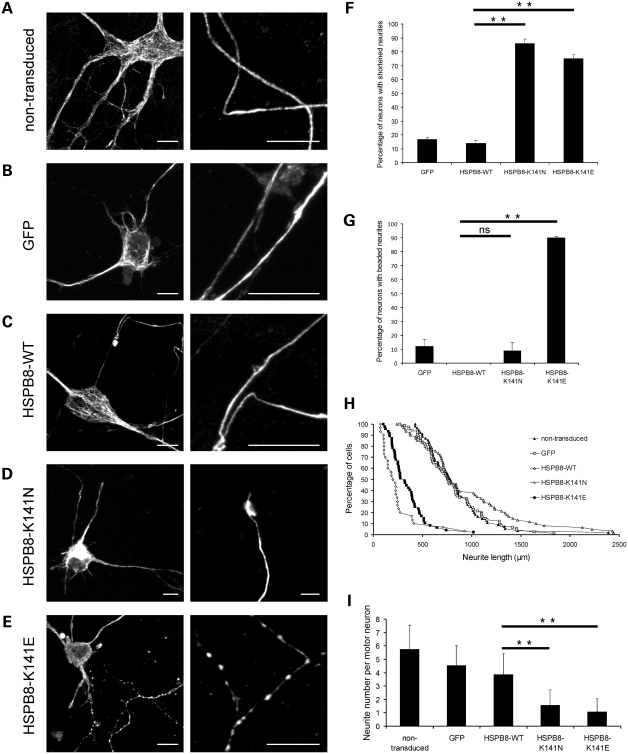
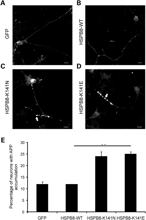
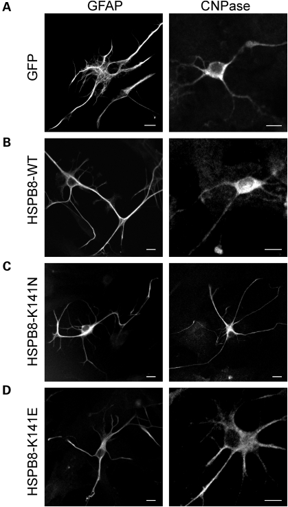
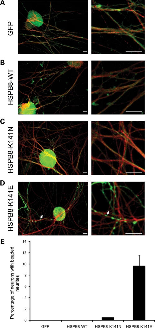
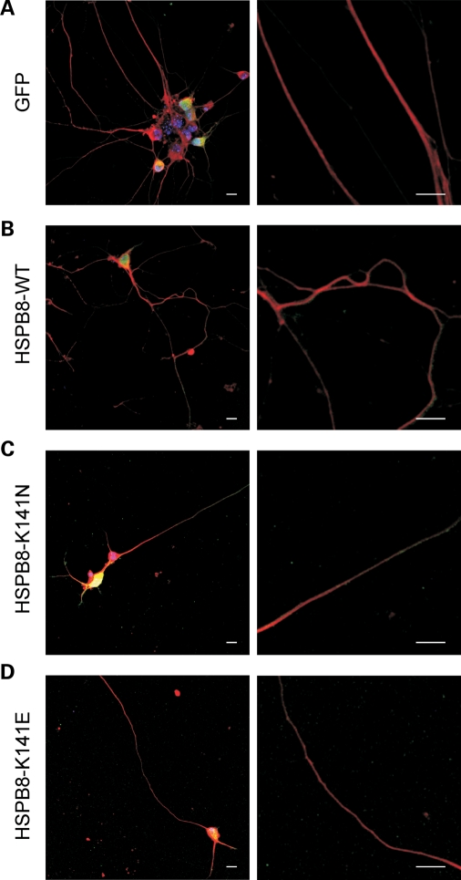
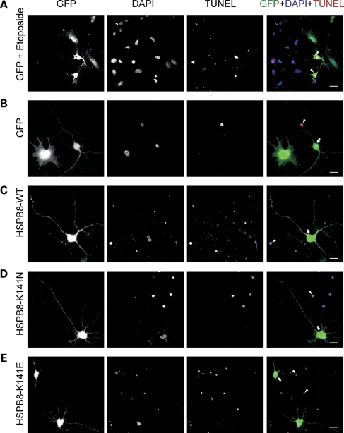
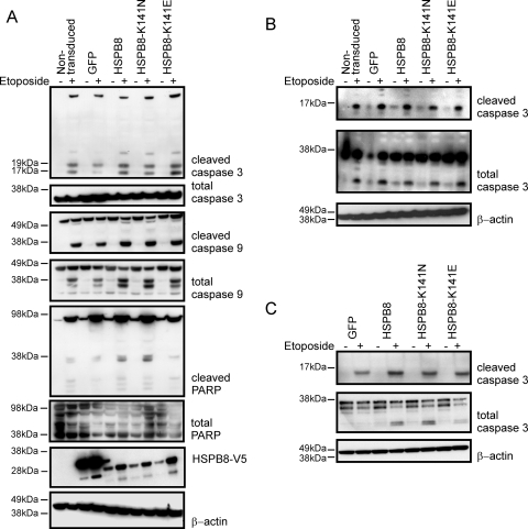
Missense mutations (K141N and K141E) in the alpha-crystallin domain of the small heat shock protein HSPB8 (HSP22) cause distal hereditary motor neuropathy (distal HMN) or Charcot-Marie-Tooth neuropathy type 2L (CMT2L). The mechanism through which mutant HSPB8 leads to a specific motor neuron disease phenotype is currently unknown. To address this question, we compared the effect of mutant HSPB8 in primary neuronal and glial cell cultures. In motor neurons, expression of both HSPB8 K141N and K141E mutations clearly resulted in neurite degeneration, as manifested by a reduction in number of neurites per cell, as well as in a reduction in average length of the neurites. Furthermore, expression of the K141E (and to a lesser extent, K141N) mutation also induced spheroids in the neurites. We did not detect any signs of apoptosis in motor neurons, showing that mutant HSPB8 resulted in neurite degeneration without inducing neuronal death. While overt in motor neurons, these phenotypes were only very mildly present in sensory neurons and completely absent in cortical neurons. Also glial cells did not show an altered phenotype upon expression of mutant HSPB8. These findings show that despite the ubiquitous presence of HSPB8, only motor neurons appear to be affected by the K141N and K141E mutations which explain the predominant motor neuron phenotype in distal HMN and CMT2L.


Figures

Figure 1.
Figure 1.
Mutant HSPB8 induces neurite abnormalities in primary motor neurons. Rat motor neurons were transduced with pLenti-GFP, pLenti-WT-HSPB8-GFP or mutant pLenti-K141N/K141E-HSPB8-GFP constructs at day in vitro 3 (DIV3) and immunostained against the non-phosphorylated epitope in neurofilament H (SMI32 antibody) at DIV7. Non-transduced motor neurons (A), motor neurons expressing native GFP (B) and WT-HSPB8 (C) show a punctate and homogenous neurofilament distribution in the motor neuron soma and normal formation of long and intact neurites, while neuritic processes of motor neurons expressing mutant HSPB8-K141N are shortened (D), and motor neurons expressing mutant HSPB8-K141E exhibited clear signs of neurite degeneration (spheroids or beaded neurites) (E). Scale bar = 10 µm. The incidence of neurite abnormalities in the motor neuron cultures were quantified by counting the proportion of neurons with abnormal (reduced or shortened) neurites (F) and with clear signs of neurite degeneration (beaded neurites) (G), by measuring neurite length distribution (H) and by counting the number of neurites per neuron (I). ns = not significant, **P-value < 0.01.
Figure 2.
Figure 2.
Motor neurons expressing mutant HSPB8 show enhanced neuritic APP accumulation. Rat motor neurons were transduced with pLenti-GFP, pLenti-WT-HSPB8-GFP or mutant pLenti-K141N/K141E-HSPB8-GFP constructs at DIV3 and immunostained using amyloid precursor protein (APP) antibody at DIV7. Motor neuron neurites expressing GFP (A) or HSPB8-WT (B) mainly show a faint and punctate appearance of APP in the neurites, similar to non-transduced neurites, while neurites of motor neuron expressing HSPB8-K141N (C) or HSPB8-K141E (D) accumulate more APP (arrow). The incidence of APP accumulation was quantified by counting the proportion of cells with strong neuritic APP accumulation (E). **P-value < 0.01. Scale bar = 10 µm.
Figure 3.
Figure 3.
Expression of mutant HSPB8 has no detectable effect on the morphology of primary glial cells. Rat glial cells were transduced with pLenti-GFP, pLenti-WT-HSPB8-GFP or mutant pLenti-K141N/K141E-HSPB8-GFP constructs at DIV3 and immunostained at DIV7 with antibodies against either GFAP (left) or 2′, 3′-cyclic nucleotide 3′-phosphodiesterase (CNPase, right). The glial cells expressing GFP (A), WT-HSPB8 (B), mutant HSPB8-K141N (C) or HSPB8-K141E (D) were morphologically similar. Scale bar = 10 µm.
Figure 4.
Figure 4.
Mutant HSPB8 induces neurite degeneration in a small proportion of primary sensory neurons. Mouse sensory neurons were transduced with pLenti-GFP, pLenti-WT-HSPB8-GFP or mutant pLenti-K141N/K141E-HSPB8-GFP constructs at DIV6 and immunostained at DIV10 using β-III tubulin antibody. Merged confocal micrographs of GFP (green) and β-III tubulin (red) are shown (A–D). No signs of neurite degeneration were found in sensory neurons expressing GFP (A), HSPB8-WT (B) and HSPB8-K141N (C), while the neurites of some cells expressing HSPB8-K141E were clearly beaded (arrow) (D). Scale bar = 10 µm. The incidence of neurite degeneration was quantified by counting the proportion of neurons with beaded neurites (E). **P-value < 0.01.
Figure 5.
Figure 5.
Cortical neurons expressing mutant HSPB8 do not show neurite abnormalities. Mouse cortical neurons were transduced with pLenti-GFP (A), pLenti-WT-HSPB8-GFP (B), mutant pLenti-K141N-HSPB8-GFP (C) or pLenti-K141E-HSPB8-GFP (D) constructs at DIV3 and immunostained with β-III tubulin at DIV7. Merged confocal micrographs of GFP (green) and β-III tubulin (red) are shown. Expression of the different HSPB8 constructs did not result in abnormal neurite formation. Scale bar = 10 µm.
Figure 6.
Figure 6.
Apoptosis analysis of motor neurons expressing mutant HSPB8. Rat motor neurons were transduced with pLenti-GFP, pLenti-WT-HSPB8-GFP or mutant pLenti-K141N/K141E-HSPB8-GFP constructs at DIV3 and analyzed for the activation of apoptosis at DIV7 with the TUNEL assay which detects DNA breaks in the nuclei of apoptotic cells. Confocal micrographs are shown of GFP expression (green in merge), nuclei of all cells (apoptotic and non-apoptotic, DAPI, blue in merge) and nuclei of apoptotic cells (TUNEL, red in merge). Treating the motor neuron cultures with 50 µm etoposide for 2 h was used as a positive control for cell death activation (A). Expression of the different constructs did not lead to detectable TUNEL staining (B–E). An additional internal positive control was provided by the TUNEL-positive nuclei resulting from dead cells always present in the primary cultures (arrowheads). Motor neurons expressing mutant K141E-HSPB8-GFP exhibit beaded or shortened neurites, but do not have TUNEL-positive nuclei (E). Arrowheads indicate TUNEL-positive cells and arrows indicate GFP or HSPB8-positive TUNEL-negative cells. Scale bar = 10 µm.
Figure 7.
Figure 7.
Expression of mutant HSPB8 does not result in increased activation of apoptosis in different neuronal and non-neuronal cell types. Activation of enzymes implicated in initiation and execution of apoptosis was examined by western blot of cleaved (17, 19 kDa) and total caspase 3 (35 kDa), cleaved (35, 37 kDa) and total caspase 9 (47 kDa) and cleaved (89 kDa) and total PARP (116 kDa) in different cell types. Stimulation with etoposide (4 h in 50 µM) was used as a positive control for apoptosis activation in the human neuronal cell line SH-SY5Y stably expressing GFP, WT-HSPB8-V5 or mutant K141N/K141E-HSPB8-V5 constructs (A), in rat primary motor neurons (B) and in rat primary glial cells (C) transduced with pLenti-GFP, pLenti-WT-HSPB8-GFP or mutant pLenti-K141N/K141E-HSPB8-GFP constructs. Expression of mutant HSPB8 did not result in increased basal levels or in increased activation after etoposide treatment of the markers for apoptosis. Normalization was performed with β-actin.

Similar articles

Cited by

References

    1. Dierick I., Irobi J., De Jonghe P., Timmerman V. Small heat shock proteins in inherited peripheral neuropathies. Ann. Med. 2005;37:413–422. doi:10.1080/07853890500296410. - DOI - PubMed
    1. Haslbeck M., Franzmann T., Weinfurtner D., Buchner J. Some like it hot: the structure and function of small heat-shock proteins. Nat. Struct. Mol. Biol. 2005;12:842–846. doi:10.1038/nsmb993. - DOI - PubMed
    1. Walter S., Buchner J. Molecular chaperones—cellular machines for protein folding. Angew. Chem. Int. Ed Engl. 2002;41:1098–1113. doi:10.1002/1521-3773(20020402)41:7<1098::AID-ANIE1098>3.0.CO;2-9. - DOI - PubMed
    1. Tang B.S., Zhao G.H., Luo W., Xia K., Cai F., Pan Q., Zhang R.X., Zhang F.F., Liu X.M., Chen B., et al. Small heat-shock protein 22 mutated in autosomal dominant Charcot-Marie-Tooth disease type 2L. Hum. Genet. 2005;116:222–224. doi:10.1007/s00439-004-1218-3. - DOI - PubMed
    1. Irobi J., Van Impe K., Seeman P., Jordanova A., Dierick I., Verpoorten N., Michalik A., De Vriendt E., Jacobs A., Van Gerwen V., et al. Hot-spot residue in small heat-shock protein 22 causes distal motor neuropathy. Nat. Genet. 2004;36:597–601. doi:10.1038/ng1328. - DOI - PubMed

Publication types

MeSH terms