. 2010 Aug;299(2):R509-20.
doi: 10.1152/ajpregu.00858.2009. Epub 2010 Jun 10.

Sepsis and glucocorticoids upregulate p300 and downregulate HDAC6 expression and activity in skeletal muscle

Affiliations

Sepsis and glucocorticoids upregulate p300 and downregulate HDAC6 expression and activity in skeletal muscle

Nima Alamdari et al. Am J Physiol Regul Integr Comp Physiol. 2010 Aug.

Abstract

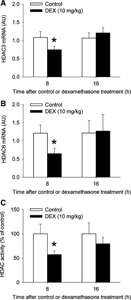
Muscle wasting during sepsis is in part regulated by glucocorticoids. In recent studies, treatment of cultured muscle cells in vitro with dexamethasone upregulated expression and activity of p300, a histone acetyl transferase (HAT), and reduced expression and activity of the histone deacetylases-3 (HDAC3) and -6, changes that favor hyperacetylation. Here, we tested the hypothesis that sepsis and glucocorticoids regulate p300 and HDAC3 and -6 in skeletal muscle in vivo. Because sepsis-induced metabolic changes are particularly pronounced in white, fast-twitch skeletal muscle, most experiments were performed in extensor digitorum longus muscles. Sepsis in rats upregulated p300 mRNA and protein levels, stimulated HAT activity, and reduced HDAC6 expression and HDAC activity. The sepsis-induced changes in p300 and HDAC expression were prevented by the glucocorticoid receptor antagonist RU38486. Treatment of rats with dexamethasone increased expression of p300 and HAT activity, reduced expression of HDAC3 and -6, and inhibited HDAC activity. Finally, treatment with the HDAC inhibitor trichostatin A resulted in increased muscle proteolysis and expression of the ubiquitin ligase atrogin-1. Taken together, our results suggest for the first time that sepsis-induced muscle wasting may be regulated by glucocorticoid-dependent hyperacetylation caused by increased p300 and reduced HDAC expression and activity. The recent development of pharmacological HDAC activators may provide a novel avenue to prevent and treat muscle wasting in sepsis and other catabolic conditions.


Figures

Fig. 1.
Fig. 1.
Sepsis in rats increases p300 expression and histone acetyl transferase (HAT) activity in skeletal muscle. A: p300 mRNA levels were determined by real-time PCR in extensor digitorum longus (EDL) muscles from sham-operated rats and rats made septic by cecal ligation and puncture (CLP). AU, arbitrary units. Results are means ± SE with n = 8 per group. *P < 0.05 vs. sham by ANOVA. B: p300 protein levels were determined by coimmunoprecipitation in EDL muscles 16 h after sham operation or CLP. Top: representative immunoblots and graph shows quantification by densitometry of 6 blots in each group. Results are means ± SE with n = 6 in each group. *P < 0.05 vs. sham by Student's t-test. C: HAT activity was measured in EDL muscles from sham-operated rats and rats made septic by CLP. Results are means ± SE with n = 8 per group. *P < 0.05 vs. sham by ANOVA.
Fig. 2.
Fig. 2.
Histone deacetylases (HDAC)3 and -6 mRNA levels in skeletal muscle during sepsis in rats. HDAC3 (A) and HDAC6 (B) mRNA levels were determined by real-time PCR in EDL muscles at different time points after sham operation or induction of sepsis by CLP in rats. Results are means ± SE with n = 8 in each group. Statistical analysis was performed using ANOVA.
Fig. 3.
Fig. 3.
Sepsis in rats reduces protein levels of HDAC6 and SIRT1 (a class III HDAC) in skeletal muscle. Protein levels of HDAC3 (A), HDAC6 (B), and SIRT1 (C) were determined by Western blot analysis of nuclear proteins. OCT1 or lamin A/C levels were determined for loading control. Representative blots are shown. Bars represent densitometric quantifications of 7 or 8 blots. Results are means ± SE. *P < 0.05 vs. sham determined by Student's t-test.
Fig. 4.
Fig. 4.
Sepsis in rats reduces HDAC activity in skeletal muscle. HDAC activity was measured as described in materials and methods in EDL muscles at different time points after sham operation or CLP. Results are means ± SE with n = 8 in each group. *P < 0.05 vs. sham by ANOVA.
Fig. 5.
Fig. 5.
Sepsis in rats increases the expression of the ubiquitin ligases atrogin-1 and muscle ring finger 1 (MuRF1) in skeletal muscle. Atrogin-1 (A) and MuRF1 (B) mRNA levels were determined by real-time PCR in EDL muscles at different time points after sham operation or CLP. Results are means ± SE with n = 8 in each group. *P < 0.05 vs. sham at the corresponding time point by ANOVA. Atrogin-1 (C) and MuRF1 (D) protein levels were determined by Western blot analysis. Representative blots are shown. Bars represent densitometric quantifications of 8 blots in each group and results are means ± SE. *P < 0.05 vs. sham by Student's t-test.
Fig. 6.
Fig. 6.
Treatment of rats with dexamethasone increases the expression of p300 and stimulates HAT activity in skeletal muscle. Levels of p300 mRNA (A) and HAT activity (B) were determined in EDL muscles 8 and 16 h after treatment of rats with 10 mg/kg of dexamethasone or vehicle (control). Results are means ± SE with n = 8 in each group. *P < 0.05 vs. contol by ANOVA.
Fig. 7.
Fig. 7.
Treatment of rats with dexamethasone reduces the expression of HDAC3 and -6 and inhibits HDAC activity in skeletal muscle. HDAC3 (A) and HDAC6 (B) mRNA levels and HDAC activity (C) were determined in EDL muscles 8 and 16 h after treatment of rats with 10 mg/kg of dexamethasone or vehicle (control). Results are means ± SE with n = 8 in each group. *P < 0.05 vs. control at the corresponding time point by ANOVA.
Fig. 8.
Fig. 8.
Treatment of rats with dexamethasone increases the expression of the ubiquitin ligases atrogin-1 and MuRF1 in skeletal muscle. Atrogin-1 (A) and MuRF1 (B) mRNA levels were determined by real-time PCR in EDL muscles 8 and 16 h after treatment of rats with 10 mg/kg of dexamethasone or vehicle (control). Results are means ± SE with n = 8 in each group. *P < 0.05 vs. control at the corresponding time point by ANOVA.
Fig. 9.
Fig. 9.
Treatment of rats with the glucocorticoid receptor antagonist RU38486 prevents sepsis-induced changes in p300 and HDAC6 expression in skeletal muscle. A: p300 mRNA levels were determined by real-time PCR in EDL muscles 16 h after sham operation or CLP. Rats were treated with 10 mg/kg of RU38486 or vehicle (control) by intraperitoneal injection 2 h before sham operation or CLP. HDAC3 (B) and HDAC6 (C) mRNA levels were determined in EDL muscles 8 h after sham operation or CLP. Rats were treated with 10 mg/kg of RU38486 or vehicle (control) by intraperitoneal injection 2 h before sham operation or CLP. Results are means ± SE with n = 8 in each group. *P < 0.05 vs. control by ANOVA.
Fig. 10.
Fig. 10.
Treatment of rats with trichostatin A (TSA) reduces HDAC activity and increases protein degradation and atrogin-1 expression in skeletal muscle. A: HDAC activity was measured in EDL muscles 2 and 4 h after treatment of rats with 10 mg/kg of TSA or vehicle (control). B: protein breakdown rates were determined by measuring tyrosine release in incubated EDL muscles 2 and 4 h after treatment with TSA or vehicle. Atrogin-1 (C) and MuRF1 (D) mRNA levels were determined by real-time PCR in EDL muscles 2 and 4 h after treatment with TSA or vehicle. Results are means ± SE with n = 6 or 7 in each group. *P < 0.05 vs. control by ANOVA.
Fig. 11.
Fig. 11.
Sepsis-induced changes in soleus muscles of p300, HDAC3, HDAC6, atrogin-1, and MuRF1 mRNA levels. The mRNA levels were determined by real-time PCR at different time points after sham operation or CLP. Results are means ± SE with n = 6–8 in each group. *P < 0.05 vs. sham by ANOVA.

Similar articles

Cited by

References

    1. Arany Z, Lebrasseur N, Morris C, Smith E, Yang W, Ma Y, Chin S, Spiegelman BM. The transcriptional coactivator PGC-1β drives the formation of oxidative type IIX fibers in skeletal muscle. Cell Metab 5: 35–46, 2007 - PubMed
    1. Avila AM, Burnett BG, Taye AA, Gabanella F, Knight MA, Hartenstein P, Cizman Z, Di Prospero NA, Pellizoni L, Fischbeck KH, Sumner CJ. Trichostatin A increases SMN expression and survival in a mouse model of spinal muscular atrophy. J Clin Invest 117: 659–671, 2007 - PMC - PubMed
    1. Bodine SC, Latres E, Baumheuter S, Lai VK, Nunez L, Clarke BA, Poueymiron WT, Panaro FJ, Na E, Dharmarajan K, Pan ZQ, Valenzuela DM, Dechiara TM, Stitt TN, Yancopoulos GD, Glass DJ. Identification of ubiquitin ligases required for skeletal muscle atrophy. Science 294: 1704–1708, 2001 - PubMed
    1. Cai D, Frantz JD, Tawa NE, Melandez PA, Oh BC, Lidov HGW, Hasselgren PO, Frontera WR, Lee J, Glass DJ, Shoelson SE. IKKβ/NF-kB activation causes severe muscle wasting in mice. Cell 119: 285–298, 2004 - PubMed
    1. Canto C, Gerhart-Hines Z, Feige JN, Lagouge M, Noriega L, Milne JC, Elliot PJ, Puigserver P, Auwerx J. AMPK regulates energy expenditure by modulating NAD+ metabolism and SIRT1 activity. Nature 458: 1056–1060, 2009 - PMC - PubMed

Publication types

MeSH terms

Substances

LinkOut - more resources