. 2010 Jun 11;328(5984):1391-4.
doi: 10.1126/science.1187703.

Exploitation of the intestinal microflora by the parasitic nematode Trichuris muris

Affiliations

Exploitation of the intestinal microflora by the parasitic nematode Trichuris muris

K S Hayes et al. Science. .

Abstract

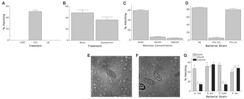
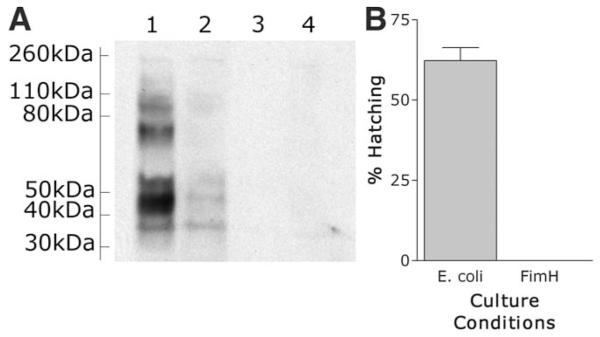
The inhabitants of the mammalian gut are not always relatively benign commensal bacteria but may also include larger and more parasitic organisms, such as worms and protozoa. At some level, all these organisms are capable of interacting with each other. We found that successful establishment of the chronically infecting parasitic nematode Trichuris muris in the large intestine of mice is dependent on microflora and coincident with modulation of the host immune response. By reducing the number of bacteria in the host animal, we significantly reduced the number of hatched T. muris eggs. Critical interactions between bacteria (microflora) and parasites (macrofauna) introduced a new dynamic to the intestinal niche, which has fundamental implications for our current concepts of intestinal homeostasis and regulation of immunity.


Figures

Fig. 1
Fig. 1
T. muris eggs are induced to hatch in vitro by contact with bacteria. (A) Hatching of T. muris eggs for 2 hours at 37°C incubated with 5-cm sections of mouse cecum, E. coli bacterial suspension, or 0.22-μm filtered (overnight) bacterial suspension. Luria-Bertani (LB) broth was used as negative control. (B) T. muris eggs were cultured with four strains of bacteria (E. coli, S. aureus, S. typhimurium, or P. aeruginosa) for 2 hours at 37°C. (C) T. muris eggs incubated with Saccharomyces cerevisiae, with or without 0.4-μm transwell or 0.22-μm filtered S. cerevisiae cultures for 2 hours at 37°C. (D) Hatching of T. muris eggs in E. coli bacterial suspension or with 0.4-μm or 3-μm transwells. (E) T. muris eggs incubated for 2 hours with E. coli at 4°C, room temperature (RT), or 37°C. Eggs that did not hatch at 4°C or room temperature were then incubated for a further 2 hours at 37°C (4/37 and RT/37, respectively). (F to H) T. muris eggs were cultured at 37°C for 1 hour with GFP-expressing bacteria, then washed in phosphate-buffered saline before further incubation for 1 hour at 37°C. (F) Light-field image; (G) fluorescence image; (H) combined image. Scale bar, 10 μm. All figures show means ± SEM, *P < 0.01, using analysis of variance (ANOVA).
Fig. 2
Fig. 2
T. muris eggs are induced to hatch in vitro after contact with type 1 fimbriae. (A) T. muris eggs were incubated for 2 hours at 37°C with E. coli preincubated for 10 min at 37° or 100°C. LB broth alone was used as a negative control. (B) T. muris eggs were incubated for 2 hours at 37°C with E. coli pretreated for 30 min with gentamicin (600 μg/ml) or untreated as a control. (C) T. muris eggs were incubated for 2 hours at 37°C with E. coli treated with 0, 50, or 100 mM mannose 30 min before and during hatching. (D) T. muris eggs were incubated for 2 hours at 37°C with wild-type E. coli (strain PK1162), FimKO E. coli, or FimKI E. coli. (E and F) T. muris eggs were incubated with FimKO (E) or FimKI (F) E. coli for 1 hour at 37°C. Scale bar, 10 μm. (G) T. muris eggs after 2 hours of incubation at 37°C with E. coli, S. aureus, S. typhimurium, or P. aeruginosa plus 0, 50, or 100 mM mannose before and during hatching. All figures show means ± SEM, *P < 0.001 using ANOVA.
Fig. 3
Fig. 3
Western blot analysis of purified FimH binding to surface egg proteins. T. muris eggs are induced to hatch in vitro after contact with FimH adhesion on type 1 fimbriae. Egg proteins were subjected to SDS–polyacrylamide gel electrophoresis under reducing conditions and electrophoretically transferred onto nitrocellulose. The migration positions of protein standards are indicated on the left. Lane 1, purified FimH and secondary antibody; lane 2, purified FimH preincubated with mannose to specifically block binding; lane 3, purified FimH only (no secondary); lane 4, secondary only.
Fig. 4
Fig. 4
T. muris eggs are induced to hatch in vivo by the presence of bacteria. (A) Worm burden at day 18 p.i.. in C57BL/6 mice treated with enrofloxacin (Baytril) from day −2 p.i. to day 18 p.i. Untreated C57BL/6 mice were used as controls. (B) IL-4 secretion by antigen restimulated MLN cells from enrofloxacin-treated and untreated control C57BL/6 mice at day 18 p.i. assessed by CBA assay. (C) IL-13 secretion. (D) IFN-γ secretion. (E) IL-6 secretion. (F) IL-17 secretion. (G) Worm burden at day 18 p.i. in SCID mice treated with enrofloxacin from day −2 p.i. to day 18 p.i. Untreated SCID mice were used as controls. (H) Worm burden at day 14 p.i. in AKR mice treated with enrofloxacin at the beginning only (day −5 to day 5) or end only (day 5 to day 13) of infection. Untreated AKR mice used as controls. All figures show means ± SEM, P < 0.01 by ANOVA.

References

    1. Ley RE, et al. Science. 2008;320:1647. published online 22 May 2008 (10.1126/science.1155725) - PMC - PubMed
    1. Turnbaugh PJ, et al. Nature. 2007;449:804. - PMC - PubMed
    1. Hooper LV, Gordon JI. Science. 2001;292:1115. - PubMed
    1. Bäckhed F, Ley RE, Sonnenburg JL, Peterson DA, Gordon JI. Science. 2005;307:1915. - PubMed
    1. Edelman SM, Kasper DL. Curr. Opin. Gastro. 2008;24:720. - PubMed

Publication types

MeSH terms