. 2010 Jul 13;107(28):12564-9.
doi: 10.1073/pnas.0914191107. Epub 2010 Jun 10.

Loss of human Greatwall results in G2 arrest and multiple mitotic defects due to deregulation of the cyclin B-Cdc2/PP2A balance

Affiliations

Loss of human Greatwall results in G2 arrest and multiple mitotic defects due to deregulation of the cyclin B-Cdc2/PP2A balance

Andrew Burgess et al. Proc Natl Acad Sci U S A. .

Abstract

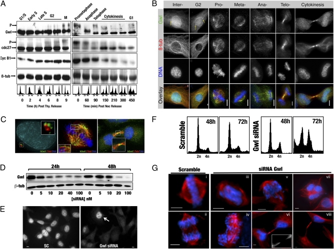
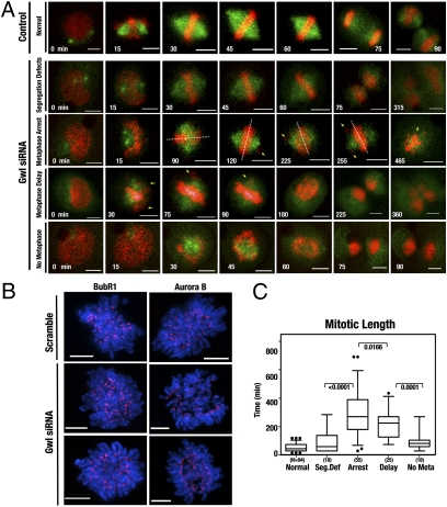
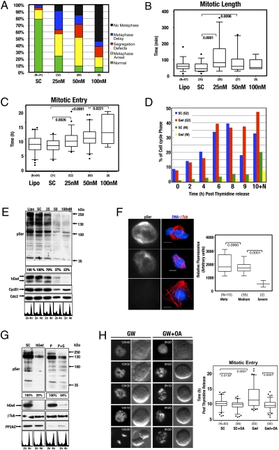
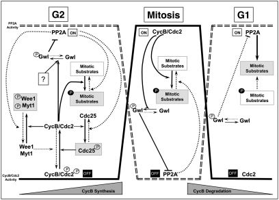
Here we show that the functional human ortholog of Greatwall protein kinase (Gwl) is the microtubule-associated serine/threonine kinase-like protein, MAST-L. This kinase promotes mitotic entry and maintenance in human cells by inhibiting protein phosphatase 2A (PP2A), a phosphatase that dephosphorylates cyclin B-Cdc2 substrates. The complete depletion of Gwl by siRNA arrests human cells in G2. When the levels of this kinase are only partially depleted, however, cells enter into mitosis with multiple defects and fail to inactivate the spindle assembly checkpoint (SAC). The ability of cells to remain arrested in mitosis by the SAC appears to be directly proportional to the amount of Gwl remaining. Thus, when Gwl is only slightly reduced, cells arrest at prometaphase. More complete depletion correlates with the premature dephosphorylation of cyclin B-Cdc2 substrates, inactivation of the SAC, and subsequent exit from mitosis with severe cytokinesis defects. These phenotypes appear to be mediated by PP2A, as they could be rescued by either a double Gwl/PP2A knockdown or by the inhibition of this phosphatase with okadaic acid. These results suggest that the balance between cyclin B-Cdc2 and PP2A must be tightly regulated for correct mitotic entry and exit and that Gwl is crucial for mediating this regulation in somatic human cells.


Conflict of interest statement

The authors declare no conflict of interest.

Figures

Fig. 1.
Fig. 1.
hGwl siRNA knockdown induces multiple mitotic defects. (A) HeLa cells were synchronized by either thymidine (Thy) or nocodazole (Noc) shake-off and released into fresh media. Samples were collected at the indicated times and processed for Western blot. Cell synchrony was confirmed by FACS. (B) HeLa cells were probed with anti-hGwl (green), anti-β-tubulin (red), and DAPI (DNA, blue). The maximum projections from 0.3-μm Z-sections are shown. Yellow arrows indicate the centrosomal localization of hGwl. (Scale bars, 5 μm.) (C) Enlargements of HeLa cells treated as per B. Cells were counterstained with γ- (interphase, red) or β-tubulin (mitotic, cytokinesis, red), hGwl (green), and DNA (blue). (D) Asynchronous HeLa cells were transfected with the indicated amounts of hGwl siRNA for 24 h and harvested immediately (24 h) or 1 d later (48 h). The levels of hGwl were analyzed by Western blot. (E) Asynchronous HeLa cells were transfected with 50 nM of siRNA and analyzed by immunofluorescence with anti-hGwl antibodies. Arrow indicates a nontransfected cell. (F) HeLa cells were treated as per D with either 50 nM of Scramble or hGwl siRNA and analyzed by FACS. (G) Asynchronous HeLa cells treated as in D were analyzed by immunofluorescence. For β-tubulin (red) and DAPI (DNA, blue). (Scale bars, 5 μm.)
Fig. 2.
Fig. 2.
hGwl knockdown promotes heterogeneity of phenotypes of varying severity in human cells. (A) HeLa cells stably expressing EB3-GFP (green) and H2B-CherryFP (red) were transfected with 50 nM of Scramble (Control) or hGwl siRNA for 24 h. After transfection, cells were synchronized with thymidine block. Movies were started 5 h after release and images were taken every 15 min. Notations indicate, time of the frame in minutes (white), lagging chromosomes (yellow arrows), metaphase plate orientation (dotted white lines), and cell death (yellow “d”). (Scale bars, 5 μm.) (B) Mitotic HeLa cells were treated with scramble or hGwl siRNA and stained with anti-Aurora B or anti-BubR1 (red) antibodies and with DAPI (DNA, blue). Shown are the deconvoluted maximum projections from 0.3-μm Z- sections. (Scale bars, 5 μm.) (C) Cells from A were separated according to their mitotic phenotype and plotted against mitotic length. Mitotic entry was determined by analyzing the first signs of DNA condensation cross-reinforced with cell rounding. Mitotic exit was scored based on chromosome segregation at anaphase or DNA decondensation. Shown are box-plots with 5 to 95% confidence intervals (black dots indicate outliers). Two-tailed unpaired Student t tests were performed to determine statistical relevance; significant P values are shown.
Fig. 3.
Fig. 3.
hGwl knockdown phenotypes can be rescued by the inactivation of PP2A. (A) Cells treated with increasing doses of hGwl siRNA were analyzed by time-lapse microscopy, as in Fig. 2A. The mitotic population was then scored on different criteria: cells that performed a correct mitosis (normal) and cells that displayed the various aberrant phenotypes (no metaphase plate, metaphase arrest, metaphase delay, and segregation defects). (B) Shown are box-plots for the mitotic length in nontransfected cells (Lipo) and cells transfected with Scramble siRNA (SC) or increasing doses of hGwl siRNA (25, 50, and 100 nM). Mitotic entry and exit was determined as in Fig.2. The small percentage of siRNA-treated cells that completed mitosis correctly was not included in the analysis, as these were likely to be untransfected cells. The total number of cells observed in each condition is listed (N). Black dots indicate outliers. (C) Similar to B except that the time of mitotic entry instead of the mitotic length was measured. (D) HeLa cells were transfected with 100 nM of siRNA and synchronized with thymidine. The number of cells at S, G2, and mitosis at the indicated times was measured by 2D-FACS (propidium Iodide versus anti-phospho Ser Cdk1 staining). In one instance, cells were released and treated with nocodazole (100 ng/mL) for 10 h to capture the mitotic population (10+N). (E) HeLa cells were transfected or not (Lipo) with scramble (SC) or increasing amounts of siRNA (25, 50, and 100 nM), synchronized by thymidine, and released into nocodazole (100 ng/mL) for 16 h. The phosphorylation of the different cyclin B-Cdc2 substrates (pSer), as well as the levels of cyclin B1, hGW, and Cdc2 were analyzed. The pSer staining was equalized against Cdc2 and the percentage of staining in each condition is indicated. (F) HeLa cells were transfected with 50 nM of siRNA, synchronized by thymidine, and 16 h later analyzed by immunofluorescence using anti-β-tubulin (red), anti-phospho Ser antibodies, and DAPI for DNA (blue). Cells were separated into the various mitotic phenotypes observed: normal metaphase (Meta), metaphase with uncongressed chromosomes or undercondensed chromatin scattered along the spindle (Medium), or cells that underwent mitotic exit (Severe). Identical acquisition exposure-time conditions were used to capture images. Scale bar, 5 μm. The intensity of staining obtained with anti-phospho Ser antibody was measured in each unaltered cell and displayed as box-plots with 5 to 95% confidence intervals. Two-tailed unpaired student t tests were performed to determine statistical relevance; significant P values are shown. (G) HeLa cells were transfected with 100 nM of human Greatwall (hGwl), PP2AC (P), or both human Greatwall and PP2AC (P + G) siRNAs. The pSer was equalized against β-tubulin. Percentage of phosphorylation of the single hGwl knockdown (hGwl) was compared with scramble siRNA knockdown (SC), whereas the percentage of phosphorylation of the double PP2AC/hGwl knockdown (P + G) was compared with single PP2AC knockdown (P). FACS analysis confirmed that the majority of cells were arrested in G2/M. (H) HeLa H2B-CherryFP cells were transfected with 100 nM of Scramble (SC) or hGwl siRNA (GW) and, 24 h later, synchronized with thymidine. Six hours after release, cells were treated (+ OA) or not (SC and Gwl) with 500 nM of OA and followed by time-lapse microscopy with images captured every 10 min. Time of mitotic entry was determined by DNA condensation and cell detachment in each cell and displayed as box-plots with 5 to 95% confidence intervals. Two-tailed unpaired student t tests were performed to determine statistical relevance; significant P values are shown.
Fig. 4.
Fig. 4.
Hypothetical model showing the different steps required for mitotic entry. Black arrows denote pathways active during mitotic entry. Dotted arrow denotes pathways that are inhibited at mitotic entry, during mitosis, or at mitotic exit.

Comment in

References

    1. Jackman MR, Pines JN. Cyclins and the G2/M transition. Cancer Surv. 1997;29:47–73. - PubMed
    1. Bollen M, Gerlich DW, Lesage B. Mitotic phosphatases: From entry guards to exit guides. Trends Cell Biol. 2009;19:531–541. - PubMed
    1. Mochida S, Ikeo S, Gannon J, Hunt T. Regulated activity of PP2A-B55 delta is crucial for controlling entry into and exit from mitosis in Xenopus egg extracts. EMBO J. 2009;28:2777–2785. - PMC - PubMed
    1. Morgan DO. Cyclin-dependent kinases: Engines, clocks, and microprocessors. Annu Rev Cell Dev Biol. 1997;13:261–291. - PubMed
    1. Glotzer M, Murray AW, Kirschner MW. Cyclin is degraded by the ubiquitin pathway. Nature. 1991;349:132–138. - PubMed

Publication types

MeSH terms