

Changes in the biomechanical response of the optic nerve head in early experimental glaucoma
- PMID: 20538991
- PMCID: PMC3061504
- DOI: 10.1167/iovs.10-5411
Changes in the biomechanical response of the optic nerve head in early experimental glaucoma
Abstract
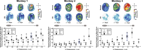
Purpose: To investigate the biomechanical response of the optic nerve head (ONH) connective tissues to IOP elevation in three pairs of monkey eyes in which one eye had early experimental glaucoma (EG).
Methods: A serial imaging technique was used to reconstruct the ONH and peripapillary sclera of three pairs of unilateral EG eyes fixed at 10 mm Hg. Eye-specific finite element models of the posterior pole were constructed with inhomogeneous material properties defined for the lamina cribrosa (LC) based on local connective tissue volume fraction (CTVF) and predominant LC beam orientation. These models were used to simulate an IOP increase from 10 to 45 mm Hg. A laminar material constant was varied to produce a range of LC displacements and scleral canal expansions, and the associated LC stress and strain were characterized.
Results: The models suggest that the LC of normal and EG eyes can deform posteriorly or anteriorly when the LC material stiffness is low or high, respectively. Scleral canal expansion was generally, but not always, reduced in EG eyes. Strains in the EG eye were similar to or lower than those in the contralateral eye for the same average LC displacement and increased when the LC was more plaint. Laminar stresses were consistently lower in the EG eye, regardless of LC stiffness.
Conclusions: Connective tissue remodeling in EG alters the biomechanical response of the LC to IOP elevation in an eye-specific manner. The models indicated that the LC tissues in EG eyes were more plaint than those in the contralateral normal eyes in two of three monkeys.
Figures


References
-
- Quigley HA, Addicks EM, Green WR, Maumenee AE. Optic nerve damage in human glaucoma. II. The site of injury and susceptibility to damage. Arch Ophthalmol. 1981;99:635–649 - PubMed
-
- Hernandez MR, Igoe F, Neufeld AH. Cell culture of the human lamina cribrosa. Invest Ophthalmol Vis Sci. 1988;29:78–89 - PubMed
-
- Clark AF, Browder SL, Steely HT, Wilson K, Cantu-Crouch D, McCartney MD. Cell biology of the human lamina cribrosa. In: Drance SM, Anderson DR. ed. Optic Nerve in Glaucoma. Amsterdam/New York: Kugler Publications; 1995:79–105
-
- Hernandez MR. The optic nerve head in glaucoma: role of astrocytes in tissue remodeling. Prog Retin Eye Res. 2000;19:297–321 - PubMed
-
- Burgoyne CF, Downs JC, Bellezza AJ, Suh JK, Hart RT. The optic nerve head as a biomechanical structure: a new paradigm for understanding the role of IOP-related stress and strain in the pathophysiology of glaucomatous optic nerve head damage. Prog Retin Eye Res. 2005;24:39–73 - PubMed
Publication types
MeSH terms
Grants and funding
LinkOut - more resources
Full Text Sources
Medical
