. 2010 Nov;51(11):5623-9.
doi: 10.1167/iovs.09-4992. Epub 2010 Jun 10.

Suppressive effects of azithromycin on zymosan-induced production of proinflammatory mediators by human corneal epithelial cells

Affiliations

Suppressive effects of azithromycin on zymosan-induced production of proinflammatory mediators by human corneal epithelial cells

De-Quan Li et al. Invest Ophthalmol Vis Sci. 2010 Nov.

Abstract

Purpose: In addition to its antibiotic effects, azithromycin has been noted to have anti-inflammatory activity, particularly in the context of microbial infections. This study was conducted to explore the suppressive effects of azithromycin on the production of proinflammatory mediators by human corneal epithelial cells (HCECs) stimulated by a fungal component, zymosan.

Methods: Primary HCECs were cultured from donor corneal limbal explants and grown to subconfluence. The cells were treated with toll-like receptor (TLR) 2 agonist zymosan (1-50 μg/mL) for 4 to 48 hours, with or without preincubation with azithromycin (1-50 μg/mL), TLR2 antibody, or NF-κB activation inhibitor quinazoline (NF-κB-I). The cells were subjected to total RNA extraction, reverse transcription (RT), and real-time PCR using gene expression assays. Cells treated for 48 hours were used for immunofluorescence staining and Western blot analysis, and their medium supernatants were collected for protein quantitation by immunobead assays.

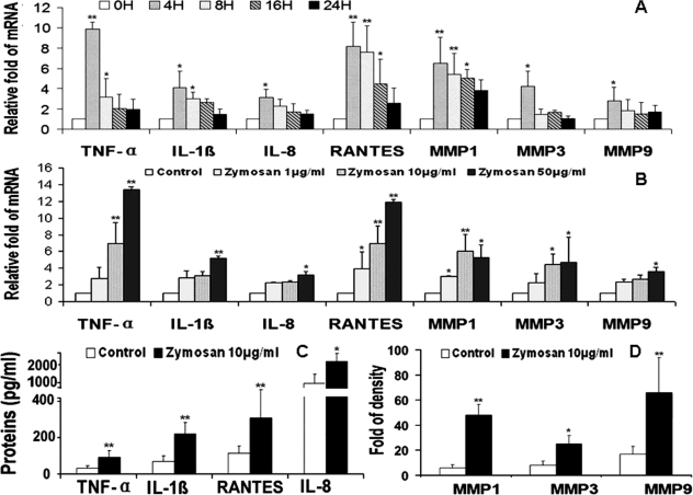
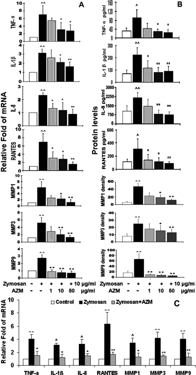
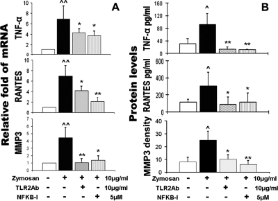
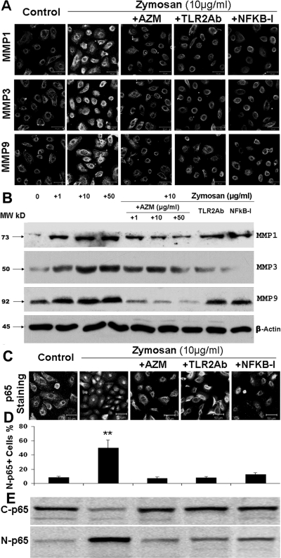
Results: The mRNA expression and protein production of proinflammatory cytokines (TNF-α and IL-1β), chemokines (IL-8 and RANTES), and matrix metalloproteinases (MMP-1, -3, and -9) by HCECs were stimulated by zymosan in a concentration-dependent manner, with peak levels noted at 4 hours. These stimulated levels of proinflammatory mediators by zymosan were significantly inhibited by TLR2 antibody, NF-κB-I, or azithromycin, which blocked zymosan-induced NF-κB activation as determined by p65 protein nuclear translocation.

Conclusions: These findings demonstrated that the fungal component zymosan induces proinflammatory responses through TLR2 and NF-κB signaling pathways, whereas azithromycin suppresses its stimulation by blocking NF-κB activation in HCECs, suggesting the potential efficacy of this antibiotic for treating ocular surface inflammatory disorders.


Figures

Figure 1.
Figure 1.
Zymosan induces proinflammatory cytokines TNF-α and IL-1β, chemokines IL-8 and RANTES, and MMP-1, -3, and -9, in HCECs. (A) The time course (0, 4, 8, 16, and 24 hours) of zymosan (10 μg/mL) on the induction of mRNA expression of these seven inflammatory mediators, evaluated by RT and real-time PCR. (B) Concentration-dependent responses of mRNA levels of these seven inflammatory mediators by HCECs exposed to zymosan (1, 10, and 50 μg/mL) for 4 hours. (C) Concentration of TNF-α, IL-1β, IL-8, and RANTES in supernatants of HCECs exposed to zymosan (10 μg/mL) for 48 hours, measured by immunobead assay. (D) Levels of MMP-1, -3, and -9 by HCECs exposed to zymosan (10 μg/mL) for 48 hours, evaluated by relative density of MMP bands in Western blot analyses. Experiments were repeated three times. Values represent mean ± SD. *P < 0.05 and **P < 0.01 compared with the control group.
Figure 2.
Figure 2.
Azithromycin concentration dependently suppressed zymosan-induced production of proinflammatory cytokines (TNF-α and IL-1β), chemokines (IL-8 and RANTES), and MMPs (MMP-1, -3, and -9) by HCECs. (A) Levels of mRNA transcripts, as evaluated by RT and real-time PCR, after stimulation by 10 μg/mL zymosan for 4 hours with or without preincubation of azithromycin (AZM; 1, 10, and 50 μg/mL) for 1 hour. (B) Protein levels, as evaluated by immunobead assay (TNF-α, IL-1β, IL-8, and RANTES) and relative density of MMP bands in Western blot analysis, after stimulation by 10 μg/mL zymosan for 48 hours in the absence and presence of azithromycin (AZM; 1, 10, and 50 μg/mL). (C) Levels of mRNA transcripts after stimulation by 10 μg/mL zymosan for 8 hours in the absence or presence of 10 μg/mL azithromycin in the last 4 hours of incubation. Experiments were repeated three times. Values represent mean ± SD. ∧P < 0.05 and ∧∧P < 0.01 compared with control group. *P < 0.05 and **P < 0.01 compared with zymosan-stimulated group.
Figure 3.
Figure 3.
TLR2 neutralizing mAb (TLR2Ab) and NF-κB activation inhibitor quinazoline (NF-κB-I) blocked the zymosan-induced production of cytokine (TNF-α), chemokine (RANTES), and MMP-3 by HCECs. (A) mRNA transcript levels, as evaluated by RT and real-time PCR, after stimulation by 10 μg/mL zymosan for 4 hours with or without preincubation of TLR2Ab (10 μg/mL) or NF-κB-I (5 μM) for 1 hour. (B) Protein levels, as evaluated by immunobead assay (TNF-α and RANTES) and relative density of MMP-3 Western blot bands in HCECs, stimulated by 10 μg/mL zymosan for 48 hours in the absence and presence of TLR2Ab (10 μg/mL) or NF-κB-I (5 μM). Experiments were repeated three times. Values represent mean ± SD. ∧P < 0.05 and ∧∧P < 0.01 compared with control group. *P < 0.05 and **P < 0.01 compared with zymosan-stimulated group.
Figure 4.
Figure 4.
Azithromycin suppresses zymosan-induced MMP production by blocking NF-κB activation. (A) Representative images of immunofluorescence staining showing stimulated production of MMP-1, -3, and -9 in HCECs exposed to 10 μg/mL zymosan for 48 hours, and suppression in the presence of TLR2Ab (10 μg/mL), NF-κB-I (5 μM), or azithromycin (AZM; 10 μg/mL). (B) Western blot analysis showing the concentration-dependent stimulation (by zymosan) and suppression (by AZM) of MMP-1, -3, and -9 with β-actin as an internal control in the condition described in (A) and (C). Representative images of immunofluorescence staining showing that NF-κB activation through p65 protein translocation from cytoplasm to nuclei was stimulated in HCECs exposed to 10 μg/mL zymosan for 4 hours and was suppressed by preincubation with TLR2Ab (10 μg/mL), NF-κB-I (5 μM), or AZM (10 μg/mL). (D) Percentages of nuclear p65-positive cells in all p65-positive cells, calculated from data in C in triplicate experiments. **P < 0.01. (E) Cells in six-well plates treated for 4 hours were subjected to cytoplasm and nuclear protein extraction for NF-κB p65 activation by Western blot analysis with 30 μg total proteins per lane. C-p65, cytoplasmic p65; N-p65, nuclear p65.

References

    1. Friedlaender MH, Protzko E. Clinical development of 1% azithromycin in DuraSite, a topical azalide anti-infective for ocular surface therapy. Clin Ophthalmol. 2007;1:3–10 - PMC - PubMed
    1. Luchs J. Efficacy of topical azithromycin ophthalmic solution 1% in the treatment of posterior blepharitis. Adv Ther. 2008;25:858–870 - PubMed
    1. Cigana C, Assael BM, Melotti P. Azithromycin selectively reduces tumor necrosis factor alpha levels in cystic fibrosis airway epithelial cells. Antimicrob Agents Chemother. 2007;51:975–981 - PMC - PubMed
    1. Cigana C, Nicolis E, Pasetto M, Assael BM, Melotti P. Anti-inflammatory effects of azithromycin in cystic fibrosis airway epithelial cells. Biochem Biophys Res Commun. 2006;350:977–982 - PubMed
    1. Ribeiro CM, Hurd H, Wu Y, et al. Azithromycin treatment alters gene expression in inflammatory, lipid metabolism, and cell cycle pathways in well-differentiated human airway epithelia. PLoS One. 2009;4:e5806. - PMC - PubMed

Publication types

MeSH terms