. 2010 Jul;11(7):522-7.
doi: 10.1038/embor.2010.81. Epub 2010 Jun 11.

Crystal structure and ligand binding of the MID domain of a eukaryotic Argonaute protein

Affiliations

Crystal structure and ligand binding of the MID domain of a eukaryotic Argonaute protein

Andreas Boland et al. EMBO Rep. 2010 Jul.

Abstract

Argonaute (AGO) proteins are core components of RNA-induced silencing complexes and have essential roles in RNA-mediated gene silencing. They are characterized by a bilobal architecture, consisting of one lobe containing the amino-terminal and PAZ domains and another containing the MID and PIWI domains. Except for the PAZ domain, structural information on eukaryotic AGO domains is not yet available. In this study, we report the crystal structure of the MID domain of the eukaryotic AGO protein QDE-2 from Neurospora crassa. This domain adopts a Rossmann-like fold and recognizes the 5'-terminal nucleotide of a guide RNA in a manner similar to its prokaryotic counterparts. The 5'-nucleotide-binding site shares common residues with a second, adjacent ligand-binding site, suggesting a mechanism for the cooperative binding of ligands to the MID domain of eukaryotic AGOs.


Conflict of interest statement

The authors declare that they have no conflict of interest.

Figures

Figure 1
Figure 1
Structure of the Neurospora crassa QDE-2 MID domain. (A) Ribbon representation of the MID domain showing the positions of two bound sulphate ions (I and II) as sticks (red: oxygen; yellow: sulphur). Residues corresponding to those proposed by Kiriakidou et al (2007) to sandwich the m7GpppN cap are shown as purple sticks. Secondary structure elements are labelled. (B) Ribbon representation of an Af MID domain in complex with the 5′-end of a guide RNA (sticks), generated from Protein Data Bank entry 2bgg (Parker et al, 2005). (C) Superposition of the Nc QDE-2 MID domain onto the Af MID domain, illustrating the conservation of the Rossmann-like fold. (D) Structure-based sequence alignment of the Nc QDE-2 MID domain with archaeal (Af, Pf) and eubacterial (Aa, Tt) homologues, as well as with eukaryotic MID domains involved in the miRNA pathway. Positions involved in coordinating the two sulphate ions are shown in green. Positions proposed by Kiriakidou et al (2007) to bind the m7GpppN cap are shown in purple. The position proposed by Djuranovic et al (2010) to effect allosteric regulation of miRNA binding is shown in pink. Invariant or highly conserved positions are marked by an asterisk. Aa, Aquifex aeolicus; Af, Archaeoglobus fulgidus; MID, middle; miRNA, micro RNA; Nc, Neurospora crassa; Pf, Pyrococcus furiosus; QDE-2, quelling deficient 2; Tt, Thermus thermophilus.
Figure 2
Figure 2
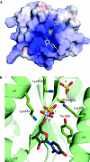
Conservation of the guide RNA 5′-nucleotide-binding pocket. (A) Electrostatic potential mapped onto the molecular surface of the Nc QDE-2 MID domain, with the two bound sulphates shown as yellow sticks. Potentials are contoured from −10 (red) to +10 kT/e (blue). The 5′-RNA nucleotide from the superposition with the Af MID domain in complex with a guide RNA (Fig 1B,C) is shown as grey sticks. (B) Close-up of the RNA 5′-end nucleotide-binding site. The superimposed 5′-uridine from the Af complex structure is shown as sticks, together with the two sulphates and important side chains from the Nc QDE-2 structure. The uridine base stacks on the aromatic Tyr 595 ring, whereas the 5′-phosphate superimposes with sulphate I. Red: oxygen; blue: nitrogen; yellow: sulphur; violet: phosphorus. Af, Archaeoglobus fulgidus; MID, middle; Nc, Neurospora crassa; QDE-2, quelling deficient 2.
Figure 3
Figure 3
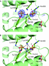
Coupled coordination of the two sulphate ion ligands. (A) Difference density for the sulphate ions. Difference density ((∣Fo∣−∣Fc∣)e(iαc)) is contoured at 3.0 sigma values over the mean, where ∣Fo∣ is the observed structure factor amplitudes and ∣Fc∣ and αc are structure factor amplitudes and phases calculated from a model prior to including the sulphates. (B) Potential hydrogen bonds of the two sulphate ions (I and II) to side-chain and main-chain atoms of the Nc QDE-2 MID domain. Relevant side chains and the main chain of strand β4 are shown as sticks. Hydrogen bonds are shown as dotted lines. Asp 603 (pink) corresponds to the aspartate proposed by Djuranovic et al (2010) to effect allosteric regulation of miRNA binding. Red: oxygen; blue: nitrogen; yellow: sulphur. MID, middle; miRNA, micro RNA; Nc, Neurospora crassa; QDE-2, quelling deficient 2.

Comment in

  • Argonaute MID domain takes centre stage.
    Faehnle CR, Joshua-Tor L. Faehnle CR, et al. EMBO Rep. 2010 Aug;11(8):564-5. doi: 10.1038/embor.2010.110. Epub 2010 Jul 16. EMBO Rep. 2010. PMID: 20634804 Free PMC article.

Similar articles

Cited by

References

    1. Adams PD et al. (2010) PHENIX: a comprehensive Python-based system for macromolecular structure solution. Acta Crystallogr D Biol Crystallogr 66: 213–221 - PMC - PubMed
    1. Broennimann C et al. (2006) The PILATUS 1M detector. J Synchrotron Radiat 13: 120–130 - PubMed
    1. Cohen SX et al. (2008) ARP/wARP and molecular replacement: the next generation. Acta Crystallogr D Biol Crystallogr 64: 49–60 - PMC - PubMed
    1. Davis IW et al. (2007) MolProbity: all-atom contacts and structure validation for proteins and nucleic acids. Nucleic Acids Res 35: W375–W383 - PMC - PubMed
    1. Djuranovic S, Zinchenko MK, Hur JK, Nahvi A, Brunelle JL, Rogers EJ, Green R (2010) Allosteric regulation of Argonaute proteins by miRNAs. Nat Struct Mol Biol 17: 144–150 - PMC - PubMed

Publication types

Associated data