. 2009 Oct;5(4):191-200.
doi: 10.4161/org.5.4.9632.

Metakaryotic stem cell lineages in organogenesis of humans and other metazoans

Affiliations

Metakaryotic stem cell lineages in organogenesis of humans and other metazoans

Elena V Gostjeva et al. Organogenesis. 2009 Oct.

Abstract

A non-eukaryotic, metakaryotic cell with large, open mouthed, bell shaped nuclei represents an important stem cell lineage in fetal/juvenile organogenesis in humans and rodents. each human bell shaped nucleus contains the diploid human DNA genome as tested by quantitative Feulgen DNA cytometry and fluorescent in situ hybridization with human pan-telomeric, pan-centromeric and chromosome specific probes. From weeks approximately 5-12 of human gestation the bell shaped nuclei are found in organ anlagen enclosed in sarcomeric tubular syncytia. Within syncytia bell shaped nuclear number increases binomially up to 16 or 32 nuclei; clusters of syncytia are regularly dispersed in organ anlagen. Syncytial bell shaped nuclei demonstrate two forms of symmetrical amitoses, facing or "kissing" bells and "stacking" bells resembling separation of two paper cups. Remarkably, DNA increase and nuclear fission occur coordinately. Importantly, syncytial bell shaped nuclei undergo asymmetrical amitoses creating organ specific ensembles of up to eight distinct closed nuclear forms, a characteristic required of a stem cell lineage. Closed nuclei emerging from bell shaped nuclei are eukaryotic as demonstrated by their subsequent increases by extra-syncytial mitoses populating the parenchyma of growing anlagen. From 9-14 weeks syncytia fragment forming single cells with bell shaped nuclei that continue to display both symmetrical and asymmetrical amitoses. These forms persist in the juvenile period and are specifically observed in bases of colonic crypts. Metakaryotic forms are found in organogenesis of humans, rats, mice and the plant Arabidopsis indicating an evolutionary origin prior to the divergence of plants and animals.

Keywords: human; metakaryote; metakaryotic cells; metazoans; organogenesis; stem cell lineage; stem cells.


Figures
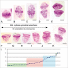
Figure 1
Figure 1
Symmetrical forms of amitosis in human fetal tissue. (A–E) “Kissing” bell symmetrical amitoses. Feulgen purple stained nuclei, arranged in order of separation of condensed DNA rings at the mouths of bell shaped nuclei. They are usually observed near midline of syncytia, but also as free single cells in earliest gestational samples, 5–7 wks [(A), brain 13 wks; (B) gut 5–7 wks; (C) brain 9 wks; (D and E) spinal cord, 9 wks]. (F–K) “Stacking cup” symmetrical amitoses. Feulgen purple stained nuclei, arranged in order of separation of condensed DNA rings at bell mouths. Usually observed throughout syncytia of 5–12 wks, but also as free single cells in post-syncytial phase, 12 weeks in fetuses to juvenile period. (L) Quantitative Feulgen estimates of DNA content (picogm DNA) as a function of average separation of condensed DNA rings at bell mouths for “stacking cup” symmetrical amitoses (as arrowed in ‘G’): Green area, separation by ∼1–2 µm; pink area, separation by ∼2–6 µm; blue area, separation ∼6–10 µm and olive area, separation 10 µm. DNA of condensed rings, ∼0.6 picogm, is doubled first in both forms of amitoses followed by ring separation [(F–I), brain 9 wks; (J) gut 5–7 wks; (K) spinal cord 9 wks]. Scale bar, 5 µm.
Figure 2
Figure 2
Bell shaped, metakaryotic nuclei in syncytia. (A) Feulgen purple stained stacking bell-to-bell symmetrical amitotic fission of bell shaped nuclei (upper) imposed over green Feulgen fluorescent image (lower) showing sarcomeric striations (arrowed) of syncytial walls (human fetal spinal cord, 9 wks). (B) Feulgen purple stained bell shaped nuclei in syncytium illustrating variety of nuclear dimensions (human fetal gut, 7 wks). (C) Green fluorescent image of a single syncytium with bell shaped nuclei (left image) and the same image merged with the image of Feulgen purple stained nuclei showing positions of the nuclei in syncytium (human fetal gut, 7 wks). Note, both “kissing cup” and “stacked cup” amitotic figures and bell shaped nucleus apparently emerging from end of syncytium. (D) Section of a Feulgen purple stained syncytium showing a portion of a larger series of eight condensed spherical nuclei (arrowed) between bell shaped nuclei interpreted as the result of synchronous asymmetrical amitoses (human fetal gut, 7 wks). Scale bar, 5 µm.
Figure 3
Figure 3
Syncytial clusters in different human fetal meta-organs. (A) Cardiac unstriated muscle at 10 weeks: Feulgen purple stained tubular syncytia (left low corner, arrows) with bell shaped nuclei in central position (zoomed image, left upper corner, 100x). Tubular syncytia are striated with bell shaped nuclei and thus distinct from differentiated cardiac unstriated muscle fibers (arrows) with closed elliptical nuclei aligned with surface of muscle fiber. (B) Skeletal striated muscle of thigh at 10 weeks: Feulgen purple stained tubular syncytia (arrows) lying in parallel with striated muscle fibers (arrowed) with sarcomeric structures with closed elliptical nuclei apparently externally associated to muscle fibers. (C) Same image as (B) merged with green Feulgen fluorescent image. Fluorescence from metakaryotic tubular syncytia is more intense than from differentiated muscle fibers. (D) Spinal cord ganglia at 9 weeks. Multiple clusters of tubular syncytia with 16 bell shaped nuclei each, green Feulgen fluorescence superimposed on purple stained Feulgen image. (E) Brain at 9 weeks. Clusters of syncytia with ∼16–32 bell shaped nuclei each stained as in (C and D). Scale bar, 100 µm.
Figure 4
Figure 4
Cells with bell shaped nuclei in post-syncytial stage in human fetal tissues. (A) Syncytial narrowing and fragmentation (arrow), brain, 13 wks. (B) Early stage of amitotic symmetrical stacking bell-to-bell nuclear fission (arrow) in narrowing syncytium (upper) and green Feulgen fluorescence image superimposed on purple stained Feulgen image (lower), brain, 13 wks. (C) Bright green Feulgen fluorescence delineates distribution of cytoplasms attached to bell shaped nuclei in small intestinal villus (11 wks). All cells with “closed” nuclear morphologies observed to date emit relatively little cytoplasmic green Feulgen fluorescence. (D) Feulgen purple stained post-syncytial bell shaped nucleus among closed nuclei (upper) and green Feulgen fluorescence superimposed on purple stained Feulgen image (lower). Note the intense Feulgen fluorescence of the post-syncytial, balloon-like cytoplasm emanating from bell shaped nucleus (human fetal colon, 12 wks). (E) Feulgen purple stained asymmetrical “bell-to-cigar” nuclear fission in post-syncytial single cell with bell shaped nucleus (left) and green Feulgen fluorescence superimposed on purple stained Feulgen image (right), small intestine, 12 wks. (F) Feulgen purple stained asymmetrical ‘bell-to-oval’ nuclear fission in post-syncytial single cell with bell shaped nucleus (left) and green Feulgen fluorescence superimposed on purple stained Feulgen image (right), small intestine, 12 wks. Scale bars, 10 µm except 40 µm in (C).
Figure 5
Figure 5
Bell shaped metakaryotic nuclei in development of animals and a plant. (A) Human gut, 7 weeks. (B) Rat, rib cage muscle, 18 days. (C) Mouse spinal cord ganglia, 16.5 days. (D) Plant Arabidopsis, embryonic stem, 1.5 days post germination. (A–D) Feulgen purple stained nuclear DNA. Note condensed, ring-like DNA at bell's mouth in all species. (E) Cluster of tubular syncytia in mouse fetal spinal cord ganglia at 14.5 days, green Feulgen fluorescence. (F) Syncytial bell shaped nuclei of mouse fetal spinal cord ganglia, 16.5 days. Green Feulgen fluorescence (left) superimposed on purple stained Feulgen image (right). (G) Symmetrical, ‘bell-to-bell’ nuclear fission in mouse spinal cord ganglia, 14.5 days. (H) Symmetrical, ‘bell-to-bell’ nuclear fission in rat rib cage muscle, 18 days. (I) Asymmetrical, ‘bell-to-sphere’ nuclear fission delineated in syncytium of a mouse spinal cord ganglia, 14.5 days, phase contrast. (J) Asymmetrical, ‘bell-to-condensed sphere’ nuclear fission in mouse myocyte cell line (Dr. J. Sherley, Boston Biomedical Research Institute, Watertown, MA). Scale bar, 5 µm, save in (D), 2 µm and in (E), 100 µm.
Figure 6
Figure 6
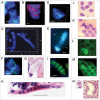
Bell shaped nuclei observed with various techniques in tissues, tumor and cell culture preparations. (A) DAPI stained DNA (blue) syncytial bell shaped nuclei in human fetal spinal cord ganglia, 8–9 wks. (B) DAPI staining (blue) and pan-telomeric FISH labeling (Cy3, red) of bell shaped nuclei in mouse fetal spinal cord ganglia tissue, 16.5 days. (C) DAPI stained syncytia with multiple bell shaped nuclei as seen in 3D imaging [Z-stack, ‘Apotome’], human fetal spinal cord ganglia, 9 wks. (D) High resolution 3D image of DAPI stained bell shaped nucleus and pan-centromeric FISH staining (FITC green), human colon adenocarcinoma, 68 yrs. (E) DAPI stained bell shaped nucleus dividing asymmetrically (bell to cigar shaped nucleus) in human colon adenocarcinoma, 68 yrs. [Arrows indicate the walls of the balloon shaped cytoplasm through which new eukaryotic nuclei migrate]. (F) DAPI stained bell shaped nucleus dividing symmetrically with segregation of pair of chromosomes 18 stained by FISH (FITC green), fetal spinal cord ganglia, 8–9 wks. (G) H&E staining of metakaryotic extra syncytial cell, colon adenocarcinoma, 68 yrs. (H) H&E staining of syncytial bell shaped nuclei, spinal cord, 8–9 wks. (I) Acridine orange stained syncytial bell shaped nuclei, spinal cord, 8–9 wks. (J and L) Feulgen stained DNA (purple) bell shaped nuclei appearing in human adenocarcinoma cell line. (K and M) Feulgen fluorescent (green) images of (J and L), respectively showing unidentified fluorescent material in cytoplasm of all cells of bell shape and with a small fraction of cells with spherical nuclei. (N) Feulgen stained bell shaped nucleus as seen in 30 microns snap frozen tissue section of human colon polyp, 27 yrs. Arrow indicates position of the nucleus relativel to aberrant crypt. Bar scales, 5 µm (A–N) except 50 µm in (H).

Similar articles

Cited by

References

    1. Gostjeva EV, Zukerberg L, Chung D, Thilly WG. Bell shaped nuclei dividing by symmetrical and asymmetrical nuclear fission have qualities of stem cells in human colonic embryogenesis and carcinogenesis. Cancer Genet Cytogenet. 2006;164:16–24. - PubMed
    1. Gostjeva EV, Thilly WG. Stem cell stages and the origins of colon cancer: a multi-disciplinary perspective. Stem Cell Rev. 2005;1:243–252. - PubMed
    1. Wilson EB. The Cell in Development and Inheritance. New York, MacMillan: 1896.
    1. Child CM. Amitosis as a factor in normal and regulatory growth. Anat Anz. 1907;30:271–297.
    1. Stough HB. Further studies of modified mitosis. J Morphol. 1935;58:221–256.

LinkOut - more resources