. 2010 Jun 7;5(6):e10988.
doi: 10.1371/journal.pone.0010988.

Cell cycle progression or translation control is not essential for vesicular stomatitis virus oncolysis of hepatocellular carcinoma

Affiliations

Cell cycle progression or translation control is not essential for vesicular stomatitis virus oncolysis of hepatocellular carcinoma

Sabrina Marozin et al. PLoS One. .

Abstract

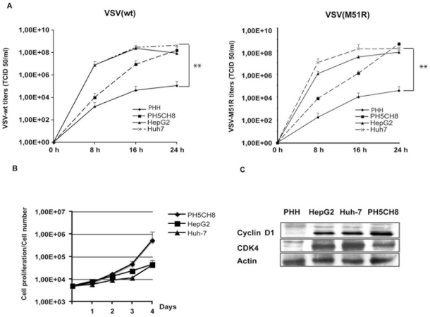
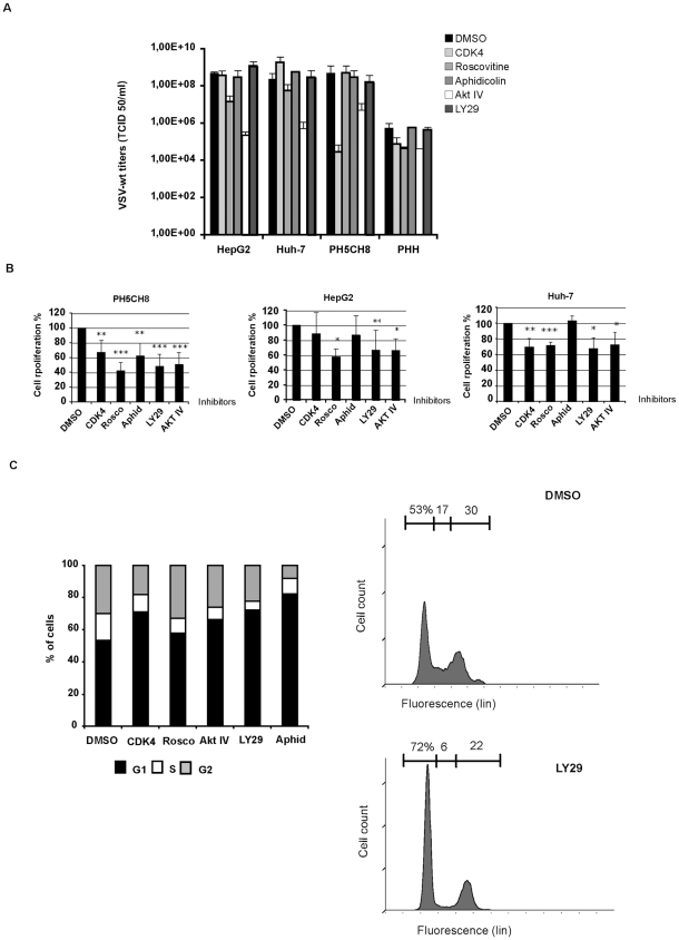
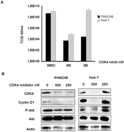
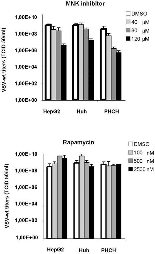
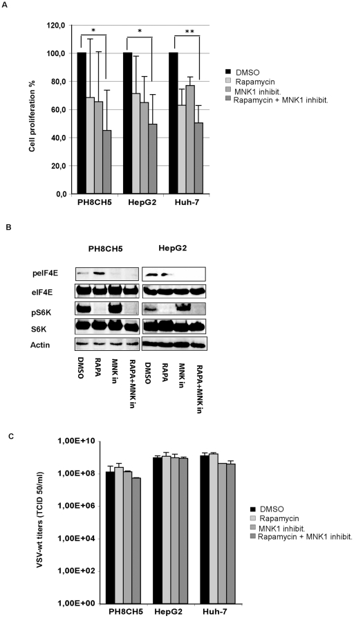
The intrinsic oncolytic specificity of vesicular stomatitis virus (VSV) is currently being exploited to develop alternative therapeutic strategies for hepatocellular carcinoma (HCC). Identifying key regulators in diverse transduction pathways that define VSV oncolysis in cancer cells represents a fundamental prerequisite to engineering more effective oncolytic viral vectors and adjusting combination therapies. After having identified defects in the signalling cascade of type I interferon induction, responsible for attenuated antiviral responses in human HCC cell lines, we have now investigated the role of cell proliferation and translation initiation. Cell cycle progression and translation initiation factors eIF4E and eIF2Bepsilon have been recently identified as key regulators of VSV permissiveness in T-lymphocytes and immortalized mouse embryonic fibroblasts, respectively. Here, we show that in HCC, decrease of cell proliferation by cell cycle inhibitors or siRNA-mediated reduction of G(1) cyclin-dependent kinase activities (CDK4) or cyclin D1 protein expression, do not significantly alter viral growth. Additionally, we demonstrate that translation initiation factors eIF4E and eIF2Bepsilon are negligible in sustaining VSV replication in HCC. Taken together, these results indicate that cellular proliferation and the initiation phase of cellular protein synthesis are not essential for successful VSV oncolysis of HCC. Moreover, our observations indicate the importance of cell-type specificity for VSV oncolysis, an important aspect to be considered in virotherapy applications in the future.


Conflict of interest statement

Competing Interests: The authors have declared that no competing interests exist.

Figures

Figure 1
Figure 1. Immortalized human hepatocytes, PH5CH8 cell line.
A) VSV growth in immortalized non-neoplastic hepatocytes (PH5CH8) was compared to HCC cell lines and primary human hepatocytes (PHH). Cells were infected with VSV-wt and the IFN-inducer mutant VSV-M51R at an MOI of 0.001 and titers were determined at different time-points post-infection as indicated. Data shown are the average of three independent experiments and error bars represent standard deviation. Significance of viral titers in PHH was calculated by comparison with titers in Huh-7 (** p<0.01). B) Proliferation of the cell lines: HepG2, Huh-7 and PH5CH8. Cells were plated at the concentration of 5×10³ cells per well, and their numbers were determined up to four days after plating by MTT proliferation assay. Data are representative of three independent experiments. C) Western blot showing the expression levels of cyclin D1 and CDK4 in HepG2, Huh-7 and PH5CH8 cells compared to PHH.
Figure 2
Figure 2. IFN system analysis in PH5CH8 cell line.
A) Fold induction of IFN-β promoter-Luciferase reporter gene in HCC cell lines (HepG2 and Huh-7), immortalized hepatocytes (PH5CH8) and primary human hepatocytes (PHH). Cells were transfected with the reporter plasmid containing the firefly luciferase gene under the control of the IFN-β promoter. At 24 hours post-transfection, cultures were stimulated by a second round of transfection with Poly (I:C) (T-pIC), Poly (I:C) was added to the medium (M-pIC), or infected with VSV-wt or VSV-M51R. IFN-luciferase activities were measured and normalized to Renilla luciferase (RL) gene used as an internal control. Significance was calculated by comparison with mock-treated cultures expressing basal firefly luciferase activity (* p<0.05; ** p<0.01; ***p<0.001). B) Interferon protection assay in PH5CH8 compared to PHH and HepG2 and Huh-7 cells as representatives for HCC. Cells were treated overnight with 500 IU/ml of universal type I interferon (IFN) or simply mock-treated. VSV-wt infection was performed at MOI of 1 and viral titers were obtained 24 hr post-infection. Titers are the mean of at least three independent experiments (* p<0.05).
Figure 3
Figure 3. Cell cycle chart and cell cycle inhibitors activity.
Scheme of cell cycle inhibitors and their specificity in blocking at a particular phase of cell cycle.
Figure 4
Figure 4. VSV replication and cell-cycle progression.
A) HCC cells (HepG2 and Huh-7) and immortalized human hepatocytes (PH5CH8) were mock-treated (DMSO) or treated with different cell-cycle inhibitors: CDK4 inhibitor (CDK4); roscovitine (Rosco); Akt inhibitor (AKT IV); Ly294002 (Ly29); Aphidicolin (Aphid). Cultures were infected with VSV-wt at an MOI of 0.1 and viral titers were determined 24 hr post-infection by TCID 50. Data represent the average of 3 independent experiments. B) MTT proliferation assay in mock-treated or cell cycle inhibitor-treated cells. C) Analysis of cell cycle phases in Huh7 cells after treatment with cycle inhibitors: CDK4 inhibitor (CDK4); roscovitine (Rosco); Akt inhibitor (AKT IV); Ly294002 (Ly29); aphidicolin (Aphid). Samples were prepared in triplicate, and representative data from three independent experiments are shown (p<0.01). Typical FACS pattern of Huh7 cells after treatment with DMSO and LY294002 and PI staining is shown.
Figure 5
Figure 5. Cell cycle inhibitors.
A broader range of concentration was tested until the appearance of cytotoxic effects. HCC cells (HepG2 and Huh-7) and immortalized human hepatocytes (PH5CH8) were mock-treated (DMSO) or treated with increasing concentrations of cell-cycle inhibitors: Roscovitine; Ly294002 and aphidicolin. Cultures were infected with VSV-wt at an MOI of 0.1 and viral titers were determined 24 hr post-infection by TCID 50. Data represent the average of 3 independent experiments.
Figure 6
Figure 6. Cell type specificity of the CDK4 inhibitor.
A) PH5CH8 and Huh-7 cells were treated with DMSO or CDK4 inhibitor at different concentrations. Infection with VSV-wt was performed at an MOI of 0.1 for 24 hr. Viral titers were determined by TCID50. Data represent the average of three independent experiments ± standard deviation. B) Protein expression of CDK4, cyclin D1 and Akt in the lysates of the above described experiment was performed by Western blot analysis.
Figure 7
Figure 7. Cell cycle arrest by siRNA.
A) Cells were transfected without siRNA, with scramble siRNA (scramb) or with siRNA against cyclin D1 or cyclin-kinase (CDK4). Forty-eight hours post-transfection cells were infected with VSV-wt at an MOI of 0.1 for 24 hours. Results show the average of at least three independent experiments. B) Mock-infected lysates from PH5CH8 and Huh-7 cells of the experiment describe above are shown for the expression of cyclin D1 and CDK4. C) FACS analysis of cell cycle arrest in Huh-7 cells upon treatment with siRNA targeting CDK4 and cyclin D1 or control siRNA (SCR). Experiments were conducted at least three times and triplicate values of one experiment are shown as representative (p<0.001).
Figure 8
Figure 8. Rapamycin activity on VSV replication.
A) HepG2, Huh-7 and PH5CH8 cells were incubated overnight with increasing concentrations of rapamycin (20, 100 and 500 nM), or in the case of the controls, DMSO was added. VSV infection was performed at an MOI of 0.1 for 24 hours in the presence of fresh inhibitor. The viral titers shown are the average of three independent experiments. B) All cell lines were treated with 50 nM of rapamycin as described above and infected with VSV-wt at an MOI of 0.1. Viral titers were determined at 8 hours post-infection. The data represent at least two independent experiments ± standard deviation. C) Cell lysates of mock- and rapamycin-treated cells were analysed by Western blot for detection of the phosphorylated forms of kinase p70S6k and eIF4E and their corresponding base-line expression. D) MTT proliferation assay in mock-treated (DMSO) and rapamycin-treated (RAPA) cultures. Data represent the mean of at least three independent experiments ± standard deviation (* p< 0.05; *** p<0.001).
Figure 9
Figure 9. VSV infection does not depend on the phosphorylation status of eIF4E.
A) Cell cultures, pre-treated with MNK1 inhibitor as described above, were infected with VSV-wt at MOI of 0.1 in the presence of fresh inhibitor. Titers were determined by TCID50 at 24 hours post-infection. Data represent the average of at least three independent experiments ± standard deviation (SD) B) Cells were pre-treated with DMSO or the MNK1 inhibitor (CGP57380, Calbiochem) at increasing concentrations for about 36 hours. Phosphorylation of eIF4E was analyzed by Western blot analyses using 100 µg of cell lysates. C) MTT assay on HCC and PH5CH8 cell lines treated with 40 µM of MNK1 inhibitor (CGP57380) are shown. Results are the average of three independent experiments ± SD (* p<0.05).
Figure 10
Figure 10. Effects of high doses of MNK inhibitor and rapamycin on VSV replication.
HCC cells (HepG2 and Huh-7) and immortalized human hepatocytes (PH5CH8) were mock-treated (DMSO) or treated with increasing concentrations of MNK inhibitor and rapamycin until the appearance of cytotoxicity. Cultures were infected with VSV-wt at an MOI of 0.1 and viral titers were determined 24 hr post-infection by TCID 50. Data represent the average of three independent experiments.
Figure 11
Figure 11. Effects of concomitant inhibition of mTOR and MNK on VSV proliferation.
Cells were mock-treated (DMSO) or treated with rapamycin at 50 nm, MNK1 inhibitor at 20 µM, alone or together as indicated. A) Cell proliferation assays were performed using the MTT assay. Representative results of at least two independent experiments are shown. B) Western blot analysis of lysates obtained by PH5CH8 and HepG2 cell lines mock-treated (DMSO) or treated with rapamycin (RAPA), MNK inhibitor (MNK in) alone or in combination (RAPA+MNK in). The levels of S6K and eIF4E phosphorylated forms were monitored after inhibitor treatment. C) Cells were infected with VSV-wt at an MOI of 0.1 for 24 hours. Viral titers represent the mean ± standard deviation of three experiments.
Figure 12
Figure 12. RNA interference assay for eIF4E and S6K.
HCC and PH5CH8 cells were transfected with siRNA for eIF4E A) or S6K B) at a concentration of 100 nM. As controls, cells were transfected in parallel with siRNA scramble or mock-transfected. At 72 hours post-transfection, cells were infected with VSV-wt using an MOI of 0.1 and viral titers were determined 8 hours post-infection. Results are the average of three independent experiments, and error bars indicate the standard deviation. Mock-infected cultures were used to control the efficiency of the mRNA silencing by Western blot analyses. Western blot analysis was performed for each experiment.
Figure 13
Figure 13. RNA interference assay for eIF2B epsilon (eIF2Bε).
A) Western blot analysis of mock-infected cultures was performed for each experiment to assess the efficiency of RNA silencing. B) HepG2, Huh-7 and PH5CH8 cells were transfected with siRNA for eIF2Bε at a concentration of 100 nM. As controls, cells were transfected in parallel with control siRNA or mock-transfected. At 72 hours post-transfection, cells were infected with VSV-wt using an MOI of 0.1 for 8 hours. Results are the average of three independent experiments and error bars indicate the standard deviation.

References

    1. Dancey JE. Therapeutic targets: MTOR and related pathways. Cancer Biol Ther. 2006;5:1065–1073. - PubMed
    1. Villanueva A, Chiang DY, Newell P, Peix J, Thung S, et al. Pivotal role of mTOR signaling in hepatocellular carcinoma. Gastroenterology. 2008;135:1972–1983, 1983e1971–1911. - PMC - PubMed
    1. Newell P, Toffanin S, Villanueva A, Chiang DY, Minguez B, et al. Ras pathway activation in hepatocellular carcinoma and anti-tumoral effect of combined sorafenib and rapamycin in vivo. J Hepatol. 2009;51:725–733. - PMC - PubMed
    1. Hallenbeck PL, Chang YN, Hay C, Golightly D, Stewart D, et al. A novel tumor-specific replication-restricted adenoviral vector for gene therapy of hepatocellular carcinoma. Hum Gene Ther. 1999;10:1721–1733. - PubMed
    1. Ebert O, Shinozaki K, Huang TG, Savontaus MJ, Garcia-Sastre A, et al. Oncolytic vesicular stomatitis virus for treatment of orthotopic hepatocellular carcinoma in immune-competent rats. Cancer Res. 2003;63:3605–3611. - PubMed

Publication types

Substances