. 2010 Jun 7;5(6):e10995.
doi: 10.1371/journal.pone.0010995.

Disrupting circadian homeostasis of sympathetic signaling promotes tumor development in mice

Affiliations

Disrupting circadian homeostasis of sympathetic signaling promotes tumor development in mice

Susie Lee et al. PLoS One. .

Abstract

Background: Cell proliferation in all rapidly renewing mammalian tissues follows a circadian rhythm that is often disrupted in advanced-stage tumors. Epidemiologic studies have revealed a clear link between disruption of circadian rhythms and cancer development in humans. Mice lacking the circadian genes Period1 and 2 (Per) or Cryptochrome1 and 2 (Cry) are deficient in cell cycle regulation and Per2 mutant mice are cancer-prone. However, it remains unclear how circadian rhythm in cell proliferation is generated in vivo and why disruption of circadian rhythm may lead to tumorigenesis.

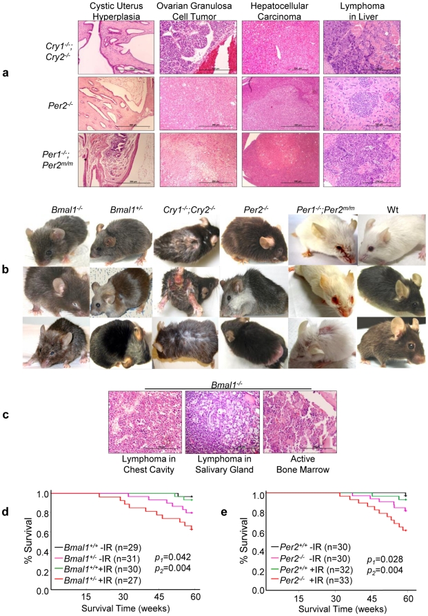
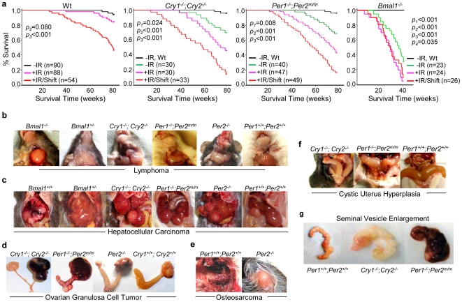
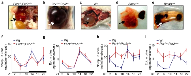
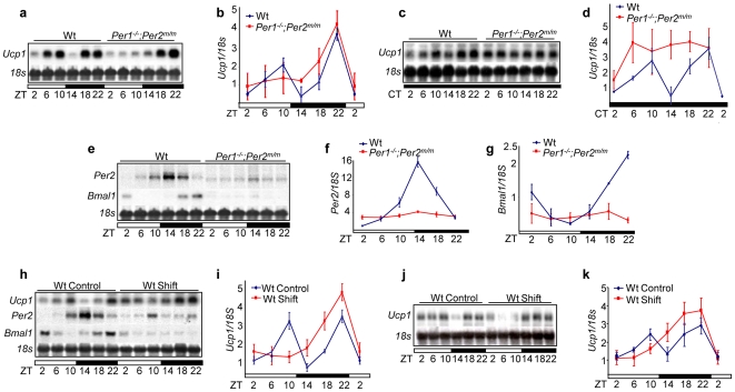
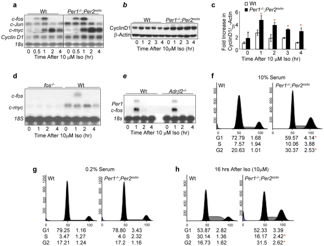
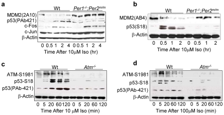
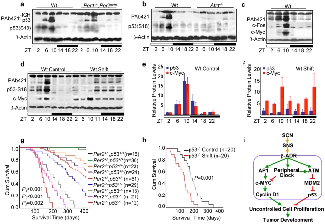
Methodology/principal findings: Mice lacking Per1 and 2, Cry1 and 2, or one copy of Bmal1, all show increased spontaneous and radiation-induced tumor development. The neoplastic growth of Per-mutant somatic cells is not controlled cell-autonomously but is dependent upon extracellular mitogenic signals. Among the circadian output pathways, the rhythmic sympathetic signaling plays a key role in the central-peripheral timing mechanism that simultaneously activates the cell cycle clock via AP1-controlled Myc induction and p53 via peripheral clock-controlled ATM activation. Jet-lag promptly desynchronizes the central clock-SNS-peripheral clock axis, abolishes the peripheral clock-dependent ATM activation, and activates myc oncogenic potential, leading to tumor development in the same organ systems in wild-type and circadian gene-mutant mice.

Conclusions/significance: Tumor suppression in vivo is a clock-controlled physiological function. The central circadian clock paces extracellular mitogenic signals that drive peripheral clock-controlled expression of key cell cycle and tumor suppressor genes to generate a circadian rhythm in cell proliferation. Frequent disruption of circadian rhythm is an important tumor promoting factor.


Conflict of interest statement

Competing Interests: The authors have declared that no competing interests exist.

Figures

Figure 1
Figure 1. Circadian Gene-mutant Mice Are Cancer-prone.
(a) Representative histological slides of cystic hyperplasia of the uterus, spontaneous ovarian and liver tumors, and lymphoma in the livers of Cry- and Per-mutant mice. (b) All mutant mouse models studied show aging phenotypes on their external appearance after exposure to a 4 Gy sublethal γ-radiation at 6 weeks of age including hair graying, alopecia, ruffled fur, skin lesions, cataracts and eye inflammation, hunchback postures, body weight changes, and sluggish activities. The ages of irradiated mice shown in (b) are: Bmal1−/− mice at 3–6 months of age, Bmal1+/− mice at 6–8 months of age, Cry1−/−;Cry2−/− mice at 3–6 months of age, Per2 −/− mice at 6–8 months of age, Per1−/−;Per2m/m mice at 6–8 months of age, and wt littermates at 6–8 months of age. (c) Representative histological slides show lymphoma in the chest cavity of a 20-week old and the salivary gland of a 36-week old irradiated Bmal1−/− mouse, and the active bone marrow in a 40 week-old irradiated Bmal1−/− mouse that also displayed aging phenotype on the external appearance. The Kaplan-Meier survival curves of (d) Bmal1+/− and (e) Per2−/− mice (−IR: untreated, +IR: irradiated, p1: untreated wt vs. untreated mutant littermates, and p2: irradiated wt vs. irradiated mutant littermates).
Figure 2
Figure 2. Disruption of Circadian Rhythm Promotes Tumor Development in Wild-type Mice.
(a) The Kaplan-Meier survival curves of wt, Cry1−/−;Cry2−/−, Per1−/−;Per2m/m and Bmal1−/− mice (−IR: untreated, +IR: irradiated, +IR/Shift: irradiated and jet-lagged, p1: untreated wt vs. untreated mutant mice, p2: untreated wt mice vs. irradiated mice, p3: untreated wt mice vs. irradiated/jet-lagged mice, and p4: untreated vs. irradiated Bmal1−/− mice). Median survival times in weeks (95% CI) are 79.7 weeks (79.2–80.1) for untreated, 78.6 weeks (77.5–79.7) for irradiated and 67.9 weeks (63.3–72.5) for irradiated/jet-lagged wt mice, 76.9 weeks (73.9–80.0) for untreated, 69.3 weeks (64.4–74.2) for irradiated and 54.9 weeks (47.8–62.0) for irradiated/jet-lagged Cry1−/−;Cry2−/− mice, 74.8 weeks (71.8–77.8) for untreated, 67.1 weeks (62.5–71.7) for irradiated and 56.1 weeks (51.0–61.2) for irradiated/jet-lagged Per1−/−;Per2m/m mice, and 35.5 weeks (30.73–40.1) for untreated, 27.3 weeks (23.3–31.4) for irradiated and 28.5 weeks (24.4–32.5) for irradiated/jet-lagged Bmal1−/− mice. Representative pictures of (b) lymphomas in the salivary glands of irradiated/jet-lagged Bmal1−/−, Bmal1+/−, Cry1−/−;Cry2−/−, Per2 −/−, Per1−/−;Per2m/m and wt (Per1+/+;Per2+/+) mice, (c) hepatocellular carcinomas in irradiated/jet-lagged Bmal1+/−, Cry1−/−;Cry2−/−, Per1−/−;Per2m/m, Per2 −/− and wt (Bmal1+/+ and Per1+/+;Per2+/+) mice, (d) ovarian granulosa cell tumors in irradiated/jet-lagged Cry1−/−;Cry2−/−, Per1−/−;Per2m/m, Per2 −/− and wt (Cry1+/+;Cry2+/+) mice, (e) osteosarcoma growing out from the spine into the chest cavity of an irradiated/jet-lagged wt (Per1+/+;Per2+/+) mouse and on the back of an irradiated/jet-lagged Per2−/− mouse, (f) severe cystic hyperplasia of the uterus in irradiated/jet-lagged Cry1−/−;Cry2−/−, Per1−/−;Per2m/m and wt (Per1+/+;Per2+/+) mice, and (g) seminal vesicles from an untreated 60-week old wt (Per1+/+;Per2+/+) mouse and age-matched irradiated/jet-lagged Cry1−/−;Cry2−/− and Per1−/−;Per2m/m mice.
Figure 3
Figure 3. Circadian Control of Sympathetic Signaling.
(a–e) Jet-lag induces fatal kidney failure in mice. The arrows indicate (a) severe cystic renal dysplasia with hydronephrosis of the remaining kidney (>20 times enlarged) in an irradiated and jet-lagged Per1−/−;Per2m/m mouse, (b) the remaining failed kidney in an irradiated and jet-lagged Cry1−/−;Cry2−/− mouse, (c) the remaining failed kidney in an irradiated and jet-lagged wt mouse, (d) the failed kidney next to the other apparently normal kidney from an untreated Bmal1−/− mouse, and (e) the remaining failed kidney with kidney stones accumulated inside in an irradiated and jet-lagged wt mouse (Bmal1+/+). (f–i) Summaries of urinary norepinephrine and epinephrine levels in wt and Per1−/−;Per2m/m mice in (f–g) 24hr LD (ZT) and (h–i) DD (CT) cycles detected from 3 to 6 independent experiments (ZT: Zeitgeber Time, with light on at ZT0 and off at ZT12; CT: Circadian Time, with CT0 as the beginning of the subjective day and CT12 as the beginning of the subjective night) (±SEM).
Figure 4
Figure 4. Central Clock-controlled Sympathetic Signaling is a Key Central-peripheral Timing Mechanism.
(a) Northern blot of Ucp1 mRNA expression in wt and Per1−/−;Per2m/m BAT in 24hr LD cycles. (b) A summary of three independent Northern blot analyses described in (a) (±SEM). (c) Ucp1 mRNA expression in wt and Per1−/−;Per2m/m BAT in 24hr DD cycles. (d) A summary of three independent Northern blot analyses described in (c) (±SEM). (e) Per2 and Bmal1 expression in wt and Per1−/−;Per2m/m BAT in 24hr LD cycles. Summaries of (f) Per2 and (g) Bmal1 mRNA expression in 24hr LD cycles from three independent experiments (±SEM). (h) Per2, Bmal1 and Ucp1 mRNA expression in BAT from untreated (Control) wt mice and wt mice treated with one cycle of jet-lag (Shift). (i) A summary of three independent Northern blot analyses described in (h) (±SEM). (j) Ucp1 mRNA expression in BAT from age-matched control wt mice and wt mice treated with 10 months of chronic jet-lag. (k) A summary of Ucp1 mRNA expression from three independent Northern blot analyses described in (j).
Figure 5
Figure 5. Hyperplastic Growth of Per-mutant Osteoblasts Is Extracellular Signal-dependent.
(a). Northern blot analysis of Ap1, c-myc and Cyclin D1 induction in 10 µM iso-treated wt and Per1−/−;Per2m/m preosteoblasts. (b) Western blot analysis of Cyclin D1 expression in 10 µM iso-treated wt and Per1−/−;Per2m/m preosteoblasts. The level of β-Actin in each sample is detected as a loading control. (c) A summary of Cyclin D1 induction from three independent Western blot analyses described in (b) (±SEM). (d) Northern blot analysis of c-myc induction by 10 µM iso in wt and c-fos −/− preosteoblasts. (e) Northern blot analysis of Per1 and c-fos induction by 10 µM iso in wt and Adrβ2 −/− preosteoblasts. The ratios of G1, S, and G2 phase cells of wt and Per1−/−;Per2m/m calvarial osteoblasts as determined by flow cytometry analysis under the normal growth condition (10% serum in culture media) (f), the serum starved condition (0.2% serum in culture media) (g), and at 16 hours after treatment with 10 µM iso (h). Per1−/−;Per2m/m osteoblasts show an increase in G2 phase in less than 16 hours after iso treatment, whereas wt osteoblasts only show an increase in S phase but not in G2 phase at the same time. The numbers below each histogram summarizes three independent flow cytometry analyses (±SEM). Asterisks indicate statistically significant differences.
Figure 6
Figure 6. Sympathetic Signaling Activates p53 via ATM.
(a) Western blot of p53, MDM2, c-FOS and c-JUN in 10 µM iso-treated wt and Per1−/−;Per2m/m osteoblasts using anti-p53 PAb421, MDM2-2A10, c-FOS and c-Jun antibodies. (b) Western blot of p53 and MDM2 in 10 µM iso-treated wt and Per1−/−;Per2m/m osteoblasts using anti-p53-S18 and MDM2-AB4 antibodies. (c) Western blot analysis of ATM-S1981 and p53 in 10 µM iso-treated wt and atm−/− osteoblasts using anti-ATM-S1981, p53-S18 and p53-PAb421 antibodies. (d) Western blot analysis of ATM and p53 in 100 µM iso-treated wt and atm−/− osteoblasts.
Figure 7
Figure 7. Circadian Expression of p53 Suppresses Tumor Development.
Western blots of p53 expression in (a) the wt and Per1−/−;Per2m/m and (b) the wt and atm−/− thymuses over a 24hr LD cycle (The PAb421 antibody also detected immunoglobulin heavy chain (IGH) in total protein extracts from the thymus, which run above p53 as a 64 kDa band). (c) Western blot of p53, c-Fos and c-Myc in thymuses of wt mice over a 24hr LD cycle. (d) A representative Western blotting of p53 and c-Myc expression in the thymus of control wt mice and wt mice treated with one cycle of jet-lag (shift). A summary of three independent Western blot analyses described in (d) for control (e) and jet-lagged (f) wt mice (±SEM). (g) The Kaplan-Meier survival curves of mice with different copies of p53 and Per2 (p1: p53+/+;Per2−/− vs. p53−/−;Per2−/− littermates, p2: p53+/+;Per2−/− vs. p53+/−;Per2−/− littermates, and p3: p53−/−;Per2+/+ vs. p53−/−;Per2−/− littermates). Median survival times in weeks (95% CI) are 55.8 weeks (52.03–59.58) for p53+/+;Per2−/− mice, 44.4 weeks (38.8–50) for p53+/−;Per2−/− mice, 28.9 weeks (25.3–32.56) for p53−/−;Per2+/+ mice, and 21 weeks (18.1–23.9) for p53−/−;Per2−/− mice. (h) The Kaplan-Meier survival curves of control and jet-lagged p53−/− mice. Median survival times in weeks (95% CI) are 27.5 weeks (23.3–31.6) for control and 17.6 weeks (14.1–21.1) for jet-lagged p53−/− mice. (i) A model for circadian control of tumor suppression.

References

    1. Hamilton T. Influence of environmental light and melatonin upon mammary tumour induction. Br J Surg. 1969;56:764–766. - PubMed
    1. Aubert C, Janiaud P, Lecalvez J. Effect of pinealectomy and melatonin on mammary tumor growth in Sprague-Dawley rats under different conditions of lighting. J Neural Transm. 1980;47:121–130. - PubMed
    1. Shah PN, Mhatre MC, Kothari LS. Effect of melatonin on mammary carcinogenesis in intact and pinealectomized rats in varying photoperiods. Cancer Res. 1984;44:3403–3407. - PubMed
    1. van den Heiligenberg S, Depres-Brummer P, Barbason H, Claustrat B, Reynes M, et al. The tumor promoting effect of constant light exposure on diethylnitrosamine-induced hepatocarcinogenesis in rats. Life Sci. 1999;64:2523–2534. - PubMed
    1. Mhatre MC, Shah PN, Juneja HS. Effect of varying photoperiods on mammary morphology, DNA synthesis, and hormone profile in female rats. J Natl Cancer Inst. 1984;72:1411–1416. - PubMed

Publication types