Review
. 2010 Aug;6(8):1431-43.
doi: 10.1039/c003913f. Epub 2010 Jun 10.

Allosteric regulation of protease activity by small molecules

Affiliations
Review

Allosteric regulation of protease activity by small molecules

Aimee Shen. Mol Biosyst. 2010 Aug.

Abstract

Proteases regulate a plethora of biological processes. Because they irreversibly cleave peptide bonds, the activity of proteases is strictly controlled. While there are many ways to regulate protease activity, an emergent mechanism is the modulation of protease function by small molecules acting at allosteric sites. This mode of regulation holds the potential to allow for the specific and temporal control of a given biological process using small molecules. These compounds also serve as useful tools for studying protein dynamics and function. This review highlights recent advances in identifying and characterizing natural and synthetic small molecule allosteric regulators of proteases and discusses their utility in studies of protease function, drug discovery and protein engineering.


Figures

Fig. 1
Fig. 1
Simplified view of allostery in proteases. Top panel: binding of an activator to an allosteric site induces a shift in the conformational equilibrium around the active site, resulting in substrate binding and cleavage. Bottom panel: binding of an inhibitor to an allosteric site causes a shift in the conformational equilibrium around the active site that no longer permits substrate binding. In both cases, there are multiple pathways (wavy lines) through which the allosteric signal is propagated. The thicker line denotes a major pathway; thinner lines represent minor pathways. It should be noted that a conformational change is not a requirement for allosteric effectors to alter protein activity.
Fig. 2
Fig. 2
Autoproteolytic activation of bacterial toxins by inositol hexakisphosphate (InsP6). (A) Structure of inositol hexakisphosphate (InsP6). Stick model and chemical structure are shown. (B) Schematic illustrating CPD-mediated autoproteolytic activation of the multidomain Clostridium sp. LGT and V. cholerae MARTX toxins. Left panel: the C-terminal end of LGTs (black squiggly line) binds to unknown cell surface receptors potentially through carbohydrate interactions. Binding results in receptor-mediated endocytosis of LGT toxins. Acidification of the endosome (H+ protons) induces a conformational change in the toxin that allows the putative translocation region (orange line) to insert into the endosomal membrane and form a pore that translocates the glucosyltransferase domain (blue triangle). The cysteine protease domain (pink circle) is also presumably translocated across the endosomal membrane. Exposure of the CPD to InsP6 (yellow jagged oval) in the eukaryotic cytosol results in protease activation. Activated CPD cleaves after a conserved Leu residue at the glucosyltransferase-CPD junction, releasing the glucosyltransferase domain into the cell, where it has improved access to Rho GTPase substrates at the plasma membrane. Right panel: the multidomain V. cholerae MARTX toxin binds to cells and is believed to autotranslocate the central effector region across the plasma membrane through conserved repeat regions (red rectangles). Exposure of the CPD to InsP6 in the eukaryotic cytosol activates the protease, which cleaves the toxin at three sites to separate effector domains. Proteolytic cleavage is necessary for optimal actin crosslinking domain activity, which induces cell rounding.
Fig. 3
Fig. 3
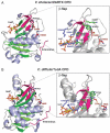
Structure of bacterial cysteine protease domains (CPDs) bound to InsP6. (A) V. cholerae MARTX CPD (PDG ID 3EEB) and (B) C. difficile TcdA CPD (PDG ID 3HO6) are shown as ribbon structures; InsP6 is shown as a stick model; the catalytic dyad Cys and His are labeled, and the side chains are shown as sticks. The N-terminus of the protein wraps around the CPD core and extends towards the active site (magenta ribbon). The β-flap structure, which bridges the InsP6 binding site and substrate binding site, is colored in pink and shown in the inset. The conserved Trp residue is shown; this residue is critical for V. cholerae CPD to communicate InsP6 binding to the active site. Residues that appear to stabilize the β-flap structure upon InsP6 binding are labeled, and the side chains are shown as green sticks. InsP6 interacting residues that may be stabilized by formation of the β-flap are indicated, and the side chains are shown as blue sticks.
Fig. 4
Fig. 4
Comparison of bacterial CPDs (Family C80) to other clan CD family members. Side and end views of (A) V. cholerae MARTX CPD (PDB ID: 3EEB), (B) C. difficile TcdA CPD (PDB ID: 3HO6), (C) Porphyromonas gingivalis gingipain R (PDB ID: ICVR), and (D) human caspase-7 (PDB ID: ISHJ) shown as ribbon structures. Proteins are rainbow colored starting with the N-terminus in blue and ending with the C-terminus in red. The catalytic Cys and His residues are labeled for orientation, and the side chains are shown as sticks.
Fig. 5
Fig. 5
A conserved mechanism for allosterically inhibiting caspases using synthetic small molecules. (A) Structure of known allosteric inhibitors. FICA and DICA inhibit the executioners caspase-3 and caspase-7, while Compound 34 inhibits the initiator caspase, caspase-1. The sulfhydryl group used to tether the compounds to Cys residues at the dimer interfaces is highlighted. (B) Comparison of three ribbon structures of mature caspase-7. Left, active site bound by Ac-DEVD-CHO inhibitor (PDB ID: 1FIJ); middle, ligand-free apoenzyme (PDB ID: 1K86); right, allosteric site-bound with DICA inhibitor (PDB ID: 1SHJ). The side chains of residues that undergo conformational rearrangements upon allosteric inhibitor binding are shown as sticks. R187, Y223, and Gly188 are shown as purple sticks; L2′ loop residues (K212-I213-P214-V215) are shown as green sticks. In the “on” state (active site-bound) structure, the allosteric dyad (R187 and Y223) are coupled through a π-cation interaction, and the L2′ loops assume an “up” position, maximizing the distance between L2′ loops. A large central cavity at the dimer interface is also apparent in the active site-bound structure. In the “off”-state structures (ligand-free and allosteric site-bound), the R187–Y223 interaction is disrupted, and the L2′ loops move in closer proximity. In the allosterically inhibited structure, the L2′ loops occlude the central cavity. (C) Surface representation of three structures of mature caspase-1 (adapted from Scheer et al.). Left, active site covalently modified by Z-VAD-FMK (PDB ID: 2HBQ); middle, ligand-free apoenzyme (PDB ID: 1SC1); right, allosteric site modified by Compound 34. The allosteric cavity at the dimer interface is indicated in red. (D) Schematic of caspase regulation following proteolytic cleavage. Once the zymogen procaspases are proteolytically activated, the caspases are in dynamic equilibrium between “zymogen-like” and “on”-state conformations in the absence of ligand. Binding of substrate (orange diamond) or modification of the active site by inhibitors shifts the equilibrium such that the protease is effectively locked in an “on-state” conformation; conversely, modification of caspases by the allosteric inhibitor (red circle) at the dimer interface traps the protease in the “off”-state similar to the zymogen. Model adapted from Scheer et al.
Fig. 6
Fig. 6
Proposed model for DD2-mediated inhibition of KSHV protease by monomer trapping. Inhibitor binding to a KSHV monomer prevents protease dimerization, effectively inhibiting enzyme activation. DD2 binds to dimerization interface residues, shifting the equilibrium towards a pre-existing folding intermediate, trapping the protease in the inactive, monomeric state. Model adapted from Shahian et al.
Fig. 7
Fig. 7
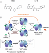
Proposed model for 1541-stimulated executioner procaspase activation. (A) Structures of 1541 and procaspase-3-specific 1541B. (B) Wolan et al. propose that procaspases are in dynamic equilibrium between “off” and “on” states, with the “off”-state being the most favored. Binding of the small molecule activator 1541 close to the active site of procaspase-3 shifts the equilibrium to an “on”-state conformation such that procaspase-3 undergoes autoproteolytic activation (black arrows). 1541 slowly stimulates removal of the prodomain and inter-subunit cleavage; the latter event is least favored. As mature caspase is produced, the autoproteolytic activation of procaspase-3 accelerates (orange arrows) due to a positive feedback loop. At high concentrations of 1541 (>30 μM), 1541 saturates the active sites and leads to inhibition. Model adapted from Wolan et al.

Similar articles

Cited by

References

    1. Turk B. Nat. Rev. Drug Discovery. 2006;5:785–799. - PubMed
    1. Bird PI, Trapani JA, Villadangos JA. Nat. Rev. Immunol. 2009;9:871–882. - PubMed
    1. del Sol A, Tsai CJ, Ma B, Nussinov R. Structure (London) 2009;17:1042–1050. - PMC - PubMed
    1. Goodey NM, Benkovic SJ. Nat. Chem. Biol. 2008;4:474–482. - PubMed
    1. Gunasekaran K, Ma B, Nussinov R. Proteins: Struct., Funct., Bioinf. 2004;57:433–443. - PubMed