

Modulation of mu rhythm desynchronization during motor imagery by transcranial direct current stimulation
- PMID: 20540721
- PMCID: PMC2898754
- DOI: 10.1186/1743-0003-7-27
Modulation of mu rhythm desynchronization during motor imagery by transcranial direct current stimulation
Abstract
Background: The mu event-related desynchronization (ERD) is supposed to reflect motor preparation and appear during motor imagery. The aim of this study is to examine the modulation of ERD with transcranial direct current stimulation (tDCS).
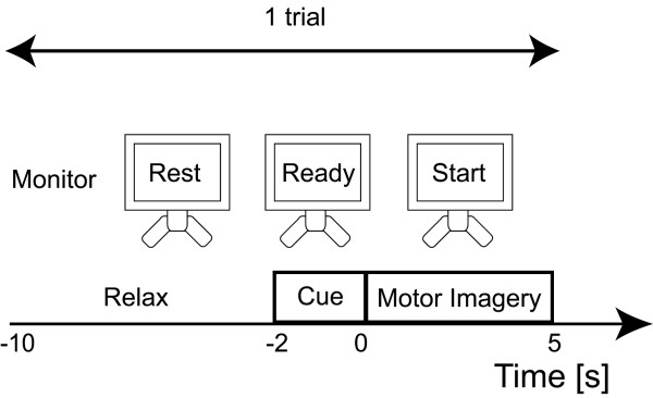
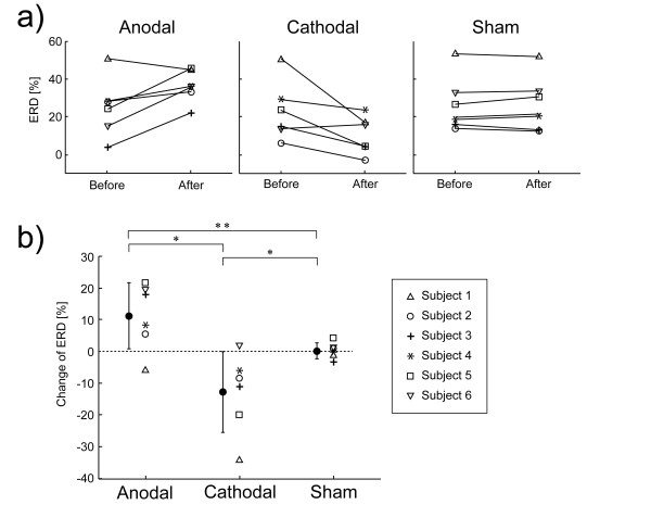
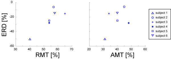
Methods: Six healthy subjects were asked to imagine their right hand grasping something after receiving a visual cue. Electroencephalograms (EEGs) were recorded near the left M1. ERD of the mu rhythm (mu ERD) by right hand motor imagery was measured. tDCS (10 min, 1 mA) was used to modulate the cortical excitability of M1. Anodal, cathodal, and sham tDCS were tested in each subject with a randomized sequence on different days. Each condition was separated from the preceding one by more than 1 week in the same subject. Before and after tDCS, mu ERD was assessed. The motor thresholds (MT) of the left M1 were also measured with transcranial magnetic stimulation.
Results: Mu ERD significantly increased after anodal stimulation, whereas it significantly decreased after cathodal stimulation. There was a significant correlation between mu ERD and MT.
Conclusions: Opposing effects on mu ERD based on the orientation of the stimulation suggest that mu ERD is affected by cortical excitability.
Figures
Similar articles
-
Transcranial direct current stimulation enhances mu rhythm desynchronization during motor imagery that depends on handedness.Laterality. 2015;20(4):453-68. doi: 10.1080/1357650X.2014.998679. Epub 2015 Jan 19. Laterality. 2015. PMID: 25599261
-
Modulation of event-related desynchronization during motor imagery with transcranial direct current stimulation (tDCS) in patients with chronic hemiparetic stroke.Exp Brain Res. 2012 Sep;221(3):263-8. doi: 10.1007/s00221-012-3166-9. Epub 2012 Jul 13. Exp Brain Res. 2012. PMID: 22791228 Clinical Trial.
-
Event-related desynchronization reflects downregulation of intracortical inhibition in human primary motor cortex.J Neurophysiol. 2013 Sep;110(5):1158-66. doi: 10.1152/jn.01092.2012. Epub 2013 Jun 12. J Neurophysiol. 2013. PMID: 23761697
-
Muscle-selective disinhibition of corticomotor representations using a motor imagery-based brain-computer interface.Neuroimage. 2018 Dec;183:597-605. doi: 10.1016/j.neuroimage.2018.08.070. Epub 2018 Aug 30. Neuroimage. 2018. PMID: 30172003
-
tDCS polarity effects in motor and cognitive domains: a meta-analytical review.Exp Brain Res. 2012 Jan;216(1):1-10. doi: 10.1007/s00221-011-2891-9. Epub 2011 Oct 12. Exp Brain Res. 2012. PMID: 21989847 Review.
Cited by
-
Brain states in freely behaving marmosets.Sleep. 2022 Aug 11;45(8):zsac106. doi: 10.1093/sleep/zsac106. Sleep. 2022. PMID: 35576961 Free PMC article.
-
Effect of tDCS stimulation of motor cortex and cerebellum on EEG classification of motor imagery and sensorimotor band power.J Neuroeng Rehabil. 2017 Apr 19;14(1):31. doi: 10.1186/s12984-017-0242-1. J Neuroeng Rehabil. 2017. PMID: 28420382 Free PMC article.
-
Post-stroke balance rehabilitation under multi-level electrotherapy: a conceptual review.Front Neurosci. 2014 Dec 15;8:403. doi: 10.3389/fnins.2014.00403. eCollection 2014. Front Neurosci. 2014. PMID: 25565937 Free PMC article.
-
Effects of neurofeedback training combined with transcranial direct current stimulation on motor imagery: A randomized controlled trial.Front Neurosci. 2023 Mar 2;17:1148336. doi: 10.3389/fnins.2023.1148336. eCollection 2023. Front Neurosci. 2023. PMID: 36937688 Free PMC article.
-
Differences in Mu rhythm when seeing grasping/motor actions in a real context versus on screens.Sci Rep. 2024 Oct 2;14(1):22921. doi: 10.1038/s41598-024-74453-x. Sci Rep. 2024. PMID: 39358411 Free PMC article.
References
Publication types
MeSH terms
LinkOut - more resources
Full Text Sources
Research Materials
