. 2010 Jun 11:7:34.
doi: 10.1186/1742-2094-7-34.

In vivo CHI3L1 (YKL-40) expression in astrocytes in acute and chronic neurological diseases

Affiliations

In vivo CHI3L1 (YKL-40) expression in astrocytes in acute and chronic neurological diseases

Dafna Bonneh-Barkay et al. J Neuroinflammation. .

Abstract

Background: CHI3L1 (YKL-40) is up-regulated in a variety of inflammatory conditions and cancers. We have previously reported elevated CHI3L1 concentration in the cerebrospinal fluid (CSF) of human and non-human primates with lentiviral encephalitis and using immunohistochemistry showed that CHI3L1 was associated with astrocytes.

Methods: In the current study CHI3L1 transcription and expression were evaluated in a variety of acute and chronic human neurological diseases.

Results: ELISA revealed significant elevation of CHI3L1 in the CSF of multiple sclerosis (MS) patients as well as mild elevation with aging. In situ hybridization (ISH) showed CHI3L1 transcription mostly associated with reactive astrocytes, that was more pronounced in inflammatory conditions like lentiviral encephalitis and MS. Comparison of CHI3L1 expression in different stages of brain infarction showed that YKL40 was abundantly expressed in astrocytes during acute phases and diminished to low levels in chronic infarcts.

Conclusions: Taken together, these findings demonstrate that CHI3L1 is induced in astrocytes in a variety of neurological diseases but that it is most abundantly associated with astrocytes in regions of inflammatory cells.


Figures

Figure 1
Figure 1
CSF CHI3L1 levels in neurodegenerative diseases and stroke. CSF samples from MS, AD, ALS, stroke and control patients were analyzed for CHI3L1 using the CHI3L1 ELISA kit. The Mann-Whitney test was used to evaluate group differences (*P < 0.05, **P < 0.001, ***P < 0.0001). CSF YKL-40 levels in MS patients were significantly elevated compared to young, age-matched controls (A). While all neurodegenerative diseases and stroke show difference from young controls, AD, ALS and stroke patients do not show significant differences when compared to older age-matched controls (B).
Figure 2
Figure 2
CHI3L1 mRNA co-localizes with CHI3L1 immunohistochemistry in SIVE. Six-μm-thick paraffin-embedded sections were hybridized with 35S-labeled RNA probe for CHI3L1 (B) followed by immunohistochemistry with for CHI3L1 (A) as described in Methods. CHI3L1 mRNA co-localizes with CHI3L1 immunohistochemistry, scale bar = 50 μm (C).
Figure 3
Figure 3
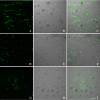
YKL-4 mRNA co-localizes with GFAP staining in SIVE. Six-μm-thick paraffin-embedded sections were hybridized with 35S-labeled RNA probe for CHI3L1 (middle panels) followed by immunohistochemistry with GFAP (A-C), NeuN (D-F) or Iba1 (G-I) as described in Methods. The right panels show the combined images of the ISH and immunohistochemistry, scale bar = 50 μm.
Figure 4
Figure 4
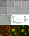
CHI3L1 mRNA in neurological diseases. Six-micrometer-thick paraffin-embedded sections from SIV encephalitis, SIV without encephalitis, AD, ALS, MS, Pick's disease, infarct and schizophrenia cases were hybridized with 35S-labeled RNA probe for CHI3L1 as described in Methods, scale bar = 100 μm (A). CHI3L1 ISH co-localized with GFAP in AD, scale bar = 20 μm (B) and brain infarction, scale bar = 50 μm (D). Higher magnification show co-localization in a single cell, scale bar = 10 μm (E). The panels to the right and on the top depict reconstructions from a confocal z-stack in xz and yz direction to confirm that CHI3L1 (green) co-localizes with GFAP (red) staining as seen in yellow (F). Five random fields from each case were captured by confocal microscopy and analyzed for CHI3L1 and GFAP pixels per field (C). Linear regression showed a positive correlation between CHI3L1 pixel count and GFAP pixel count (r2 = 0.5637; black circle SIVE, white circle SIV, black star MS, white triangle infarct, black diamond ALS, crossed square AD, gray circle Pick's disease, white circle Schizophrenia).
Figure 5
Figure 5
CHI3L1 transcription in acute, subacute and chronic infarcts. Six-micrometer-thick paraffin-embedded sections from acute (A), subacute (B) and chronic infarct (C) were hybridized with 35S-labeled RNA probe for CHI3L1 as described in Methods. Representative fields from acute, subacute and chronic infarcts were captured by confocal microscopy, scale bar = 50 μm.

References

    1. Johansen JS. Studies on serum YKL-40 as a biomarker in diseases with inflammation, tissue remodelling, fibroses and cancer. Dan Med Bull. 2006;53:172–209. - PubMed
    1. Jensen BV, Johansen JS, Price PA. High levels of serum HER-2/neu and YKL-40 independently reflect aggressiveness of metastatic breast cancer. Clin Cancer Res. 2003;9:4423–4434. - PubMed
    1. Cintin C, Johansen JS, Christensen IJ, Price PA, Sorensen S, Nielsen HJ. High serum YKL-40 level after surgery for colorectal carcinoma is related to short survival. Cancer. 2002;95:267–274. doi: 10.1002/cncr.10644. - DOI - PubMed
    1. Brasso K, Christensen IJ, Johansen JS, Teisner B, Garnero P, Price PA, Iversen P. Prognostic value of PINP, bone alkaline phosphatase, CTX-I, and YKL-40 in patients with metastatic prostate carcinoma. Prostate. 2006;66:503–513. doi: 10.1002/pros.20311. - DOI - PubMed
    1. Hormigo A, Gu B, Karimi S, Riedel E, Panageas KS, Edgar MA, Tanwar MK, Rao JS, Fleisher M, DeAngelis LM, Holland EC. YKL-40 and matrix metalloproteinase-9 as potential serum biomarkers for patients with high-grade gliomas. Clin Cancer Res. 2006;12:5698–5704. doi: 10.1158/1078-0432.CCR-06-0181. - DOI - PubMed

Publication types

MeSH terms