

Centrosome clustering and cyclin D1 gene amplification in double minutes are common events in chromosomal unstable bladder tumors
- PMID: 20540739
- PMCID: PMC2906479
- DOI: 10.1186/1471-2407-10-280
Centrosome clustering and cyclin D1 gene amplification in double minutes are common events in chromosomal unstable bladder tumors
Abstract
Background: Aneuploidy, centrosome abnormalities and gene amplification are hallmarks of chromosome instability (CIN) in cancer. Yet there are no studies of the in vivo behavior of these phenomena within the same bladder tumor.
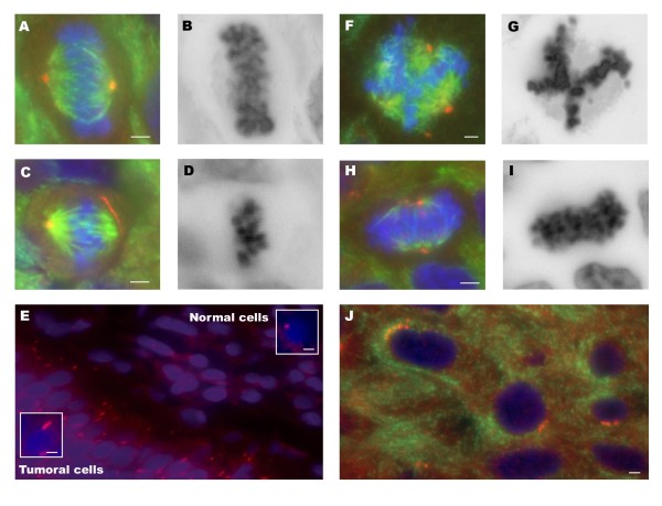
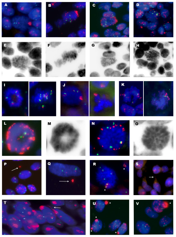
Methods: Twenty-one paraffin-embedded bladder tumors were analyzed by conventional comparative genome hybridization and fluorescence in situ hybridization (FISH) with a cyclin D1 gene (CCND1)/centromere 11 dual-color probe. Immunofluorescent staining of alpha, beta and gamma tubulin was also performed.
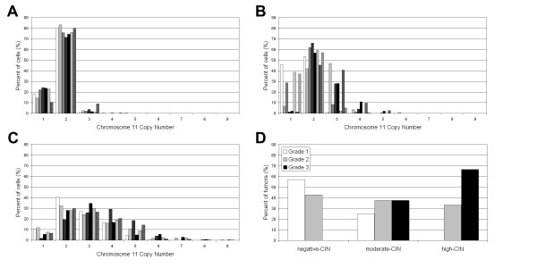
Results: Based on the CIN index, defined as the percentage of cells not displaying the modal number for chromosome 11, tumors were classified as CIN-negative and CIN-positive. Fourteen out of 21 tumors were considered CIN-positive. All T1G3 tumors were included in the CIN-positive group whereas the majority of Ta samples were classified as CIN-negative tumors. Centrosome clustering was observed in six out of 12 CIN-positive tumors analyzed. CCND1 amplification in homogeneously staining regions was present in six out of 14 CIN-positive tumors; three of them also showed amplification of this gene in double minutes.
Conclusions: Complex in vivo behavior of CCND1 amplicon in bladder tumor cells has been demonstrated by accurate FISH analysis on paraffin-embedded tumors. Positive correlation between high heterogeneity, centrosome abnormalities and CCND1 amplification was found in T1G3 bladder carcinomas. This is the first study to provide insights into the coexistence of CCND1 amplification in homogeneously staining regions and double minutes in primary bladder tumors. It is noteworthy that those patients whose tumors showed double minutes had a significantly shorter overall survival rate (p < 0.001).
Figures
Similar articles
-
CCND1/CyclinD1 status in metastasizing bladder cancer: a prognosticator and predictor of chemotherapeutic response.Mod Pathol. 2014 Jan;27(1):87-95. doi: 10.1038/modpathol.2013.125. Epub 2013 Jul 26. Mod Pathol. 2014. PMID: 23887292
-
Overexpression of polo-like kinase 1 (PLK1) and chromosomal instability in bladder cancer.Oncology. 2006;70(3):231-7. doi: 10.1159/000094416. Epub 2006 Jul 6. Oncology. 2006. PMID: 16837776
-
Centrosome hyperamplification and chromosomal instability in bladder cancer.Eur Urol. 2003 May;43(5):505-15. doi: 10.1016/s0302-2838(03)00056-3. Eur Urol. 2003. PMID: 12705995
-
Centrosome Aberrations as Drivers of Chromosomal Instability in Breast Cancer.Endocrinology. 2021 Dec 1;162(12):bqab208. doi: 10.1210/endocr/bqab208. Endocrinology. 2021. PMID: 34606589 Free PMC article. Review.
-
Deregulation of the centrosome cycle and the origin of chromosomal instability in cancer.Adv Exp Med Biol. 2005;570:393-421. doi: 10.1007/1-4020-3764-3_14. Adv Exp Med Biol. 2005. PMID: 18727509 Review.
Cited by
-
Extrachromosomal Circular DNA: A New Target in Cancer.Front Oncol. 2022 Apr 14;12:814504. doi: 10.3389/fonc.2022.814504. eCollection 2022. Front Oncol. 2022. PMID: 35494014 Free PMC article. Review.
-
Genetic instability persists in non-neoplastic urothelial cells from patients with a history of urothelial cell carcinoma.PLoS One. 2014 Jan 22;9(1):e86162. doi: 10.1371/journal.pone.0086162. eCollection 2014. PLoS One. 2014. PMID: 24465937 Free PMC article.
-
Extrachromosomal driver mutations in glioblastoma and low-grade glioma.Nat Commun. 2014 Dec 4;5:5690. doi: 10.1038/ncomms6690. Nat Commun. 2014. PMID: 25471132 Free PMC article.
-
The Renaissance of CDK Inhibitors in Breast Cancer Therapy: An Update on Clinical Trials and Therapy Resistance.Cancers (Basel). 2022 Nov 1;14(21):5388. doi: 10.3390/cancers14215388. Cancers (Basel). 2022. PMID: 36358806 Free PMC article. Review.
-
Centrosome amplification: a quantifiable cancer cell trait with prognostic value in solid malignancies.Cancer Metastasis Rev. 2021 Mar;40(1):319-339. doi: 10.1007/s10555-020-09937-z. Epub 2020 Oct 26. Cancer Metastasis Rev. 2021. PMID: 33106971 Free PMC article. Review.
References
-
- Bringuier PP, Tamimi Y, Schuuring E, Schalken J. Expression of cyclin D1 and EMS1 in bladder tumours; relationship with chromosome 11q13 amplification. Oncogene. 1996;12:1747–1753. - PubMed
Publication types
MeSH terms
Substances
LinkOut - more resources
Full Text Sources
Medical
Research Materials
