

Structural definition and substrate specificity of the S28 protease family: the crystal structure of human prolylcarboxypeptidase
- PMID: 20540760
- PMCID: PMC2893456
- DOI: 10.1186/1472-6807-10-16
Structural definition and substrate specificity of the S28 protease family: the crystal structure of human prolylcarboxypeptidase
Abstract
Background: The unique S28 family of proteases is comprised of the carboxypeptidase PRCP and the aminopeptidase DPP7. The structural basis of the different substrate specificities of the two enzymes is not understood nor has the structure of the S28 fold been described.
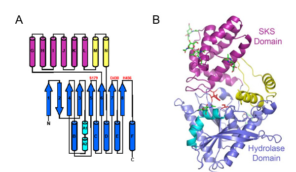
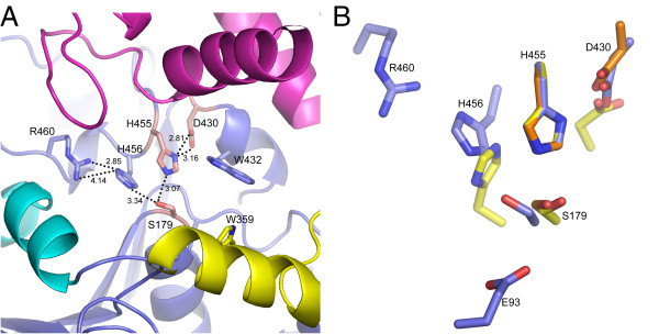
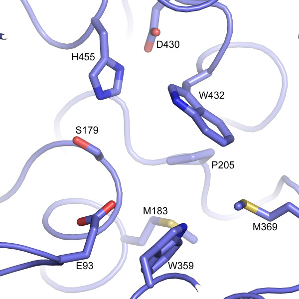
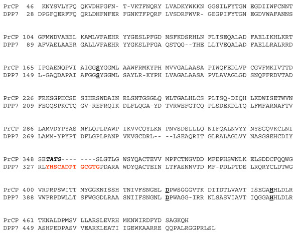
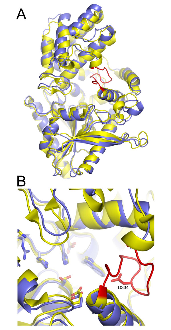
Results: The experimentally phased 2.8 A crystal structure is presented for human PRCP. PRCP contains an alpha/beta hydrolase domain harboring the catalytic Asp-His-Ser triad and a novel helical structural domain that caps the active site. Structural comparisons with prolylendopeptidase and DPP4 identify the S1 proline binding site of PRCP. A structure-based alignment with the previously undescribed structure of DPP7 illuminates the mechanism of orthogonal substrate specificity of PRCP and DPP7. PRCP has an extended active-site cleft that can accommodate proline substrates with multiple N-terminal residues. In contrast, the substrate binding groove of DPP7 is occluded by a short amino-acid insertion unique to DPP7 that creates a truncated active site selective for dipeptidyl proteolysis of N-terminal substrates.
Conclusion: The results define the structure of the S28 family of proteases, provide the structural basis of PRCP and DPP7 substrate specificity and enable the rational design of selective PRCP modulators.
Figures


Similar articles
-
Highly selective hydrolysis of kinins by recombinant prolylcarboxypeptidase.Biochem Biophys Res Commun. 2011 Feb 18;405(3):338-43. doi: 10.1016/j.bbrc.2010.12.036. Epub 2010 Dec 16. Biochem Biophys Res Commun. 2011. PMID: 21167814 Free PMC article.
-
S28 peptidases: lessons from a seemingly 'dysfunctional' family of two.BMC Biol. 2010 Jun 28;8:87. doi: 10.1186/1741-7007-8-87. BMC Biol. 2010. PMID: 20598110 Free PMC article.
-
Structures of human DPP7 reveal the molecular basis of specific inhibition and the architectural diversity of proline-specific peptidases.PLoS One. 2012;7(8):e43019. doi: 10.1371/journal.pone.0043019. Epub 2012 Aug 29. PLoS One. 2012. PMID: 22952628 Free PMC article.
-
Crystal structure and substrate recognition mechanism of the prolyl endoprotease PEP from Aspergillus niger.Biochem Biophys Res Commun. 2022 Feb 5;591:76-81. doi: 10.1016/j.bbrc.2021.12.114. Epub 2022 Jan 1. Biochem Biophys Res Commun. 2022. PMID: 34999257
-
Carboxypeptidase Y: structural basis for protein sorting and catalytic triad.J Biochem. 1999 Jul;126(1):1-6. doi: 10.1093/oxfordjournals.jbchem.a022408. J Biochem. 1999. PMID: 10393313 Review.
Cited by
-
Cloning and expression of a novel prolyl endopeptidase from Aspergillus oryzae and its application in beer stabilization.J Ind Microbiol Biotechnol. 2015 Feb;42(2):263-72. doi: 10.1007/s10295-014-1571-8. Epub 2014 Dec 30. J Ind Microbiol Biotechnol. 2015. PMID: 25547787
-
Highly selective hydrolysis of kinins by recombinant prolylcarboxypeptidase.Biochem Biophys Res Commun. 2011 Feb 18;405(3):338-43. doi: 10.1016/j.bbrc.2010.12.036. Epub 2010 Dec 16. Biochem Biophys Res Commun. 2011. PMID: 21167814 Free PMC article.
-
Prolylcarboxypeptidase Mitigates Myocardial Ischemia/Reperfusion Injury by Stabilizing Mitophagy.Front Cell Dev Biol. 2020 Oct 22;8:584933. doi: 10.3389/fcell.2020.584933. eCollection 2020. Front Cell Dev Biol. 2020. PMID: 33195231 Free PMC article.
-
A high-throughput, multiplexed assay for superfamily-wide profiling of enzyme activity.Nat Chem Biol. 2014 Aug;10(8):656-63. doi: 10.1038/nchembio.1578. Epub 2014 Jul 6. Nat Chem Biol. 2014. PMID: 24997602 Free PMC article.
-
The metabolic serine hydrolases and their functions in mammalian physiology and disease.Chem Rev. 2011 Oct 12;111(10):6022-63. doi: 10.1021/cr200075y. Epub 2011 Jun 23. Chem Rev. 2011. PMID: 21696217 Free PMC article. Review. No abstract available.
References
-
- Araki H, Li Y, Yamamoto Y, Haneda M, Nishi K, Kikkawa R, Ohkubo I. Purification, molecular cloning, and immunohistochemical localization of dipeptidyl peptidase II from the rat kidney and its identity with quiescent cell proline dipeptidase. J Biochem. 2001;129:279–288. - PubMed
-
- Chiravuri M, Schmitz T, Yardley K, Underwood R, Dayal Y, Huber BT. A novel apoptotic pathway in quiescent lymphocytes identified by inhibition of a post-proline cleaving aminodipeptidase: A candidate target protease, quiescent cell proline dipeptidase. J Immunol. 1999;163:3092–3099. - PubMed
-
- McDonald JK, Leibach FH, Grindeland RE, Ellis S. Purification of dipeptidyl aminopeptidase II (dipeptidyl arylamidase II) of the anterior pituitary gland. Peptidase and dipeptide esterase activities. J Biol Chem. 1968;243:4143–4150. - PubMed
MeSH terms
Substances
LinkOut - more resources
Full Text Sources
Other Literature Sources
Molecular Biology Databases
Miscellaneous
