. 2010 Jun 11:10:65.
doi: 10.1186/1471-213X-10-65.

An in vivo RNA interference screen identifies gene networks controlling Drosophila melanogaster blood cell homeostasis

Affiliations

An in vivo RNA interference screen identifies gene networks controlling Drosophila melanogaster blood cell homeostasis

Amélie Avet-Rochex et al. BMC Dev Biol. .

Abstract

Background: In metazoans, the hematopoietic system plays a key role both in normal development and in defense of the organism. In Drosophila, the cellular immune response involves three types of blood cells: plasmatocytes, crystal cells and lamellocytes. This last cell type is barely present in healthy larvae, but its production is strongly induced upon wasp parasitization or in mutant contexts affecting larval blood cell homeostasis. Notably, several zygotic mutations leading to melanotic mass (or "tumor") formation in larvae have been associated to the deregulated differentiation of lamellocytes. To gain further insights into the gene regulatory network and the mechanisms controlling larval blood cell homeostasis, we conducted a tissue-specific loss of function screen using hemocyte-specific Gal4 drivers and UAS-dsRNA transgenic lines.

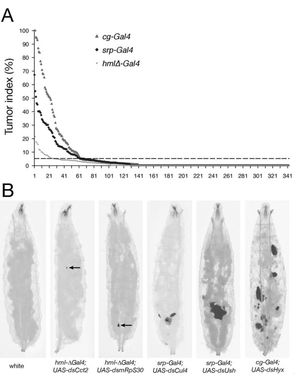
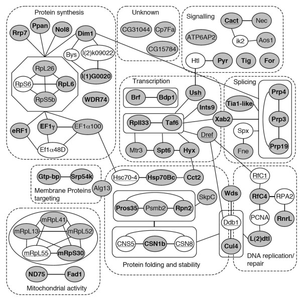
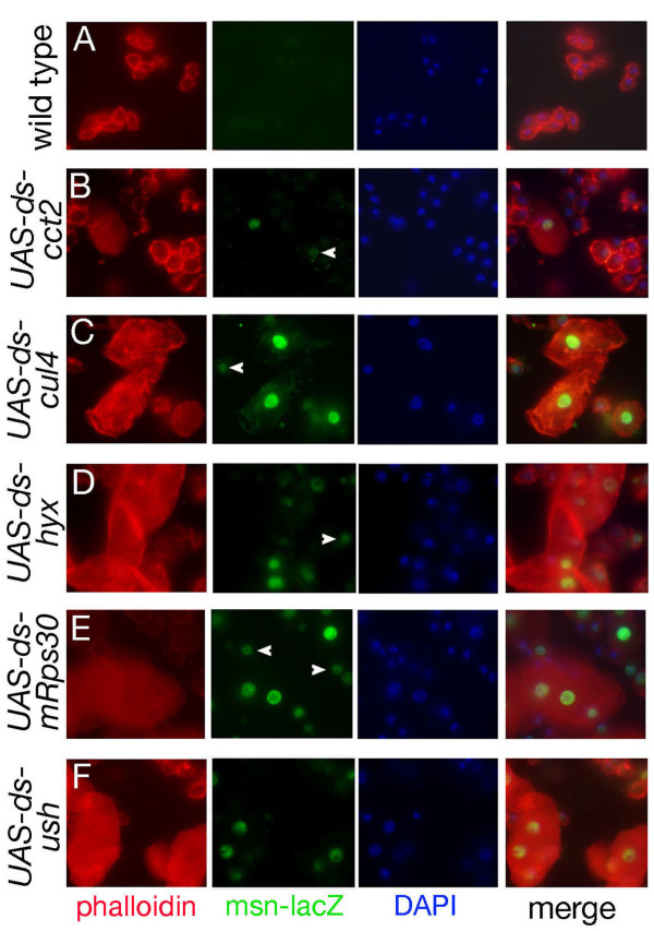
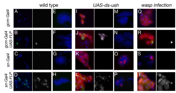
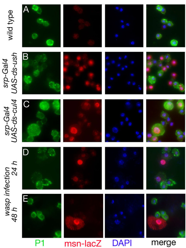
Results: By targeting around 10% of the Drosophila genes, this in vivo RNA interference screen allowed us to recover 59 melanotic tumor suppressor genes. In line with previous studies, we show that melanotic tumor formation is associated with the precocious differentiation of stem-cell like blood progenitors in the larval hematopoietic organ (the lymph gland) and the spurious differentiation of lamellocytes. We also find that melanotic tumor formation can be elicited by defects either in the fat body, the embryo-derived hemocytes or the lymph gland. In addition, we provide a definitive confirmation that lymph gland is not the only source of lamellocytes as embryo-derived plasmatocytes can differentiate into lamellocytes either upon wasp infection or upon loss of function of the Friend of GATA cofactor U-shaped.

Conclusions: In this study, we identify 55 genes whose function had not been linked to blood cell development or function before in Drosophila. Moreover our analyses reveal an unanticipated plasticity of embryo-derived plasmatocytes, thereby shedding new light on blood cell lineage relationship, and pinpoint the Friend of GATA transcription cofactor U-shaped as a key regulator of the plasmatocyte to lamellocyte transformation.


Figures

Figure 1
Figure 1
An RNAi screen for melanotic suppressor genes. (A) Distribution of tumor indices for the 342 UAS-dsRNA lines retested with the three drivers (hmlΔ-Gal4, srp-Gal4, cg-Gal4). For each of the three driver lines, the candidates are classified by decreasing tumor index (% of larvae carrying at least one melanotic nodule). The dotted line indicates the 5% threshold that we used to select 96 candidates for secondary validations. (B) Example of melanotic mass mutant phenotypes recovered in the screen. Third instar larvae are shown with their corresponding genotypes. Small melanotic masses are indicated by an arrow. Note the dissociation of the fat body in the cg-Gal4; UAS-ds-hyx larva.
Figure 2
Figure 2
An interaction network of the melanotic suppressors. The 59 confirmed melanotic suppressors identified in the screen are depicted as grey nodes together with their (inferred) name. Additional factors not identified in the screen but linking two or more melanotic suppressors are represented as white nodes. Factors previously associated with melanotic mass development and/or lamellocyte differentiation are underlined. Candidates that were confirmed using non-overlapping dsRNA are indicated in bold. The different melanotic suppressors are grouped in 9 categories (dashed lines) based on GO annotation and data mining. Factors belonging to a well-defined molecular complex are boxed together. Physical or functional interactions between the different factors are represented by edges.
Figure 3
Figure 3
Blood cell phenotypes associated to melanotic mass formation. Blood smears from third instar larvae carrying the msn-lacZ transgene and expressing UAS-dsRNA targeting the indicated gene under the control of srp-Gal4. Hemocyte actin cytoskeleton was visualized using phalloidin (red) and expression of the lamellocyte marker msn-lacZ was revealed by fluorescent immunolabeling against β-Gal (green). Nuclei were stained with DAPI. Arrowheads indicate atypical hemocytes that express β-Gal but do not exhibit the large flattened morphology of lamellocytes.
Figure 4
Figure 4
Lymph gland differentiation status. Expression of the lamellocyte differentiation marker α-ps4 (A-F) or of the prohemocyte marker tepIV (G-L) was revealed by in situ hybridization on lymph glands from early third instar larvae expressing UAS-dsRNA targeting the indicated gene under the control of srp-Gal4.
Figure 5
Figure 5
Fate of embryonic blood cells. (A-T) Blood smears (A-D, I-L and Q-T) and lymph glands (E-H and M-P) of third instar larvae. The Act > FRT > CD2 > FRT > GAL4 cassette and the UAS-FLP and UAS-GFP transgenes were used to permanently label the cells that express sn-Gal4 or gcm-Gal4. Immunostaining against GFP (green, also displayed in white on right panel of each blood smear) was used to monitor gcm-Gal4, UAS-GFP or sn-Gal4, UAS-GFP expression. In situ hybridization against α-ps4 (red) was used to reveal lamellocyte differentiation. Nuclei were counterstained with DAPI. GFP labeling alone is shown to the right. (A-H) larvae raised in wild-type conditions, (I-P) larvae carrying a UAS-dsRNA transgene against ush, (Q-T) larvae submitted to parasitization by L. boulardi. (A, E, I, M, Q) gcm-Gal4, UAS-GFP; Act > FRT > CD2 > FRT > GAL4; (B, F, J, N R) UAS-FLP; gcm-Gal4, UAS-GFP; Act > FRT > CD2 > FRT > GAL4; (C, G, K, O, S) sn-Gal4, UAS-GFP; Act > FRT > CD2 > FRT > GAL4; (D, H, L, P, T) UAS-FLP; sn-Gal4, UAS-GFP; Act > FRT > CD2 > FRT > GAL4;
Figure 6
Figure 6
Plasmatocyte and lamellocyte relationships. (A-E) Double fluorescent immunostainings on blood smears from early third instar larvae showing the expression of the plasmatocyte specific marker P1 (green) and of the lamellocyte specific marker msn-lacZ (red). Nuclei were counterstained with DAPI (blue). (A) wild type larvae, (B-C) larvae expressing dsRNA against ush (B) or cul4 (C) under the control of srp-Gal4, (D-E): larvae infected by L. boulardi, 24 h (D) or 48 h (E) after parasitization.

Similar articles

Cited by

References

    1. Hartenstein V. Blood cells and blood cell development in the animal kingdom. Annu Rev Cell Dev Biol. 2006;22:677–712. doi: 10.1146/annurev.cellbio.22.010605.093317. - DOI - PubMed
    1. Kondo M, Wagers AJ, Manz MG, Prohaska SS, Scherer DC, Beilhack GF, Shizuru JA, Weissman IL. Biology of hematopoietic stem cells and progenitors: implications for clinical application. Annu Rev Immunol. 2003;21:759–806. doi: 10.1146/annurev.immunol.21.120601.141007. - DOI - PubMed
    1. Crozatier M, Meister M. Drosophila haematopoiesis. Cell Microbiol. 2007;9:1117–1126. doi: 10.1111/j.1462-5822.2007.00930.x. - DOI - PubMed
    1. Tepass U, Fessler LI, Aziz A, Hartenstein V. Embryonic origin of hemocytes and their relationship to cell death in Drosophila. Development. 1994;120:1829–1837. - PubMed
    1. Lanot R, Zachary D, Holder F, Meister M. Postembryonic hematopoiesis in Drosophila. Dev Biol. 2001;230:243–257. doi: 10.1006/dbio.2000.0123. - DOI - PubMed

Publication types

LinkOut - more resources