

Computerized assessment of breast lesion malignancy using DCE-MRI robustness study on two independent clinical datasets from two manufacturers
- PMID: 20540907
- PMCID: PMC2907891
- DOI: 10.1016/j.acra.2010.03.007
Computerized assessment of breast lesion malignancy using DCE-MRI robustness study on two independent clinical datasets from two manufacturers
Abstract
Rationale and objectives: To conduct a preclinical evaluation of the robustness of our computerized system for breast lesion characterization on two breast magnetic resonance imaging (MRI) databases that were acquired using scanners from two different manufacturers.
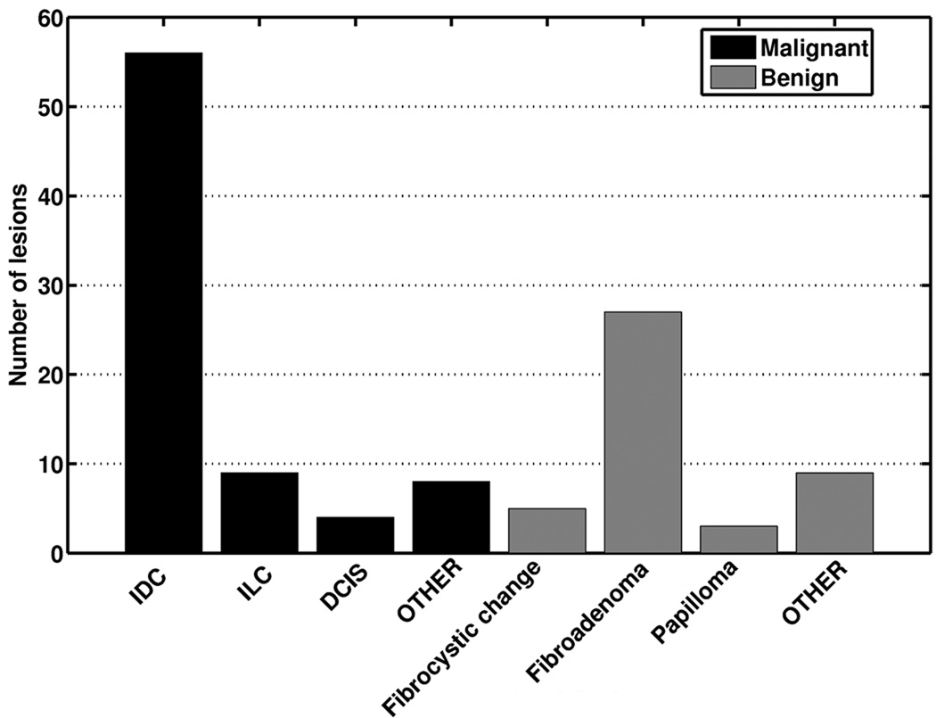
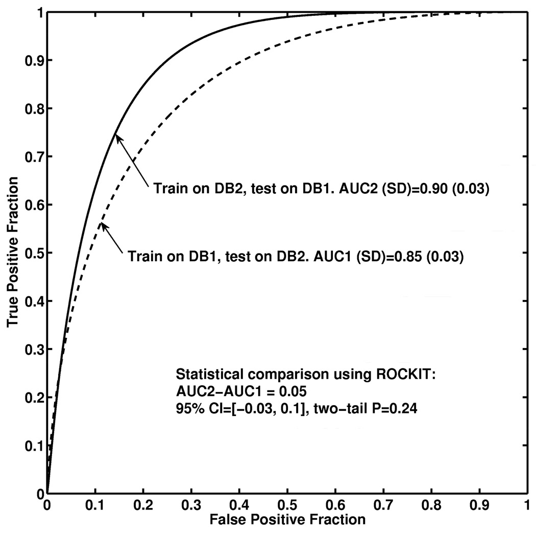
Materials and methods: Two clinical breast MRI databases were acquired from a Siemens scanner and a GE scanner, which shared similar imaging protocols and retrospectively collected under an institutional review board-approved protocol. In our computerized analysis system, after a breast lesion is identified by the radiologist, the computer performs automatic lesion segmentation and feature extraction and outputs an estimated probability of malignancy. We used a Bayesian neural network with automatic relevance determination for joint feature selection and classification. To evaluate the robustness of our classification system, we first used Database 1 for feature selection and classifier training, and Database 2 to test the trained classifier. Then, we exchanged the two datasets and repeated the process. Area under the receiver operating characteristic curve (AUC) was used as a performance figure of merit in the task of distinguishing between malignant and benign lesions.
Results: We obtained an AUC of 0.85 (approximate 95% confidence interval [CI] 0.79-0.91) for (a) feature selection and classifier training using Database 1 and testing on Database 2; and an AUC of 0.90 (approximate 95% CI 0.84-0.96) for (b) feature selection and classifier training using Database 2 and testing on Database 1. We failed to observe statistical significance for the difference AUC of 0.05 between the two database conditions (P = .24; 95% confidence interval -0.03, 0.1).
Conclusion: These results demonstrate the robustness of our computerized classification system in the task of distinguishing between malignant and benign breast lesions on dynamic contrast-enhanced (DCE) MRI images from two manufacturers. Our study showed the feasibility of developing a computerized classification system that is robust across different scanners.
2010 AUR. All rights reserved.
Figures


Similar articles
-
Multimodality computer-aided breast cancer diagnosis with FFDM and DCE-MRI.Acad Radiol. 2010 Sep;17(9):1158-67. doi: 10.1016/j.acra.2010.04.015. Acad Radiol. 2010. PMID: 20692620 Free PMC article.
-
A computerized global MR image feature analysis scheme to assist diagnosis of breast cancer: a preliminary assessment.Eur J Radiol. 2014 Jul;83(7):1086-1091. doi: 10.1016/j.ejrad.2014.03.014. Epub 2014 Mar 22. Eur J Radiol. 2014. PMID: 24743001 Free PMC article.
-
Multilevel analysis of spatiotemporal association features for differentiation of tumor enhancement patterns in breast DCE-MRI.Med Phys. 2010 Aug;37(8):3940-56. doi: 10.1118/1.3446799. Med Phys. 2010. PMID: 20879557
-
Quantitative analysis of lesion morphology and texture features for diagnostic prediction in breast MRI.Acad Radiol. 2008 Dec;15(12):1513-25. doi: 10.1016/j.acra.2008.06.005. Acad Radiol. 2008. PMID: 19000868 Free PMC article.
-
Automatic identification and classification of characteristic kinetic curves of breast lesions on DCE-MRI.Med Phys. 2006 Aug;33(8):2878-87. doi: 10.1118/1.2210568. Med Phys. 2006. PMID: 16964864
Cited by
-
Radiomics methodology for breast cancer diagnosis using multiparametric magnetic resonance imaging.J Med Imaging (Bellingham). 2020 Jul;7(4):044502. doi: 10.1117/1.JMI.7.4.044502. Epub 2020 Aug 24. J Med Imaging (Bellingham). 2020. PMID: 32864390 Free PMC article.
-
Clinical Artificial Intelligence Applications: Breast Imaging.Radiol Clin North Am. 2021 Nov;59(6):1027-1043. doi: 10.1016/j.rcl.2021.07.010. Radiol Clin North Am. 2021. PMID: 34689871 Free PMC article. Review.
-
Enhancing physicians' radiology diagnostics of COVID-19's effects on lung health by leveraging artificial intelligence.Front Bioeng Biotechnol. 2023 Apr 20;11:1010679. doi: 10.3389/fbioe.2023.1010679. eCollection 2023. Front Bioeng Biotechnol. 2023. PMID: 37152658 Free PMC article.
-
Quantitative MRI radiomics in the prediction of molecular classifications of breast cancer subtypes in the TCGA/TCIA data set.NPJ Breast Cancer. 2016;2:16012-. doi: 10.1038/npjbcancer.2016.12. Epub 2016 May 11. NPJ Breast Cancer. 2016. PMID: 27853751 Free PMC article.
-
Evaluation of Kinetic Entropy of Breast Masses Initially Found on MRI using Whole-lesion Curve Distribution Data: Comparison with the Standard Kinetic Analysis.Eur Radiol. 2015 Aug;25(8):2470-8. doi: 10.1007/s00330-015-3635-1. Epub 2015 Feb 20. Eur Radiol. 2015. PMID: 25698353
References
-
- Kuhl CK. Current status of breast MR imaging. Part 1. Technical issues. Radiology. 2007;244:356–378. - PubMed
-
- Kuhl CK. Current status of breast MR imaging. Part 2. Clinical applications. Radiology. 2007;244:672–691. - PubMed
-
- Lehman CD, Gatsonis C, Kuhl CK, Hendrick RE, Pisano ED, Hanna L, Peacock S, Smazal SF, Maki DD, Julian TB, DePeri ER, Bluemke DA, Schnall MD ACRIN Trial 6667 Investigators Group. MRI evaluation of the contralateral breast in women with recently diagnosed breast cancer. N Engl J Med. 2007;356:1295–1303. - PubMed
-
- Saslow D, Boetes C, Burke W, Harms S, Leach MO, Lehman CD, Morris E, Pisano E, Schnall M, Sener S, Smith RA, Warner E, Yaffe M, Andrews KS, Russell CA American Cancer Society Breast Cancer Advisory Group. American Cancer Society guidelines for breast screening with MRI as an adjunct to mammography. CA Cancer J Clin. 2007;57:75–89. - PubMed
-
- DeMartini W, Lehman C, Partridge S. Breast MRI for Cancer Detection and Characterization: A Review of Evidence-Based Clinical Applications. Acad Radiol. 2008;15:408–416. - PubMed
Publication types
MeSH terms
Grants and funding
LinkOut - more resources
Full Text Sources
Medical
