

Determinants of myosin II cortical localization during cytokinesis
- PMID: 20541410
- PMCID: PMC2930192
- DOI: 10.1016/j.cub.2010.04.058
Determinants of myosin II cortical localization during cytokinesis
Abstract
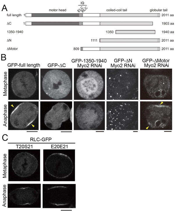
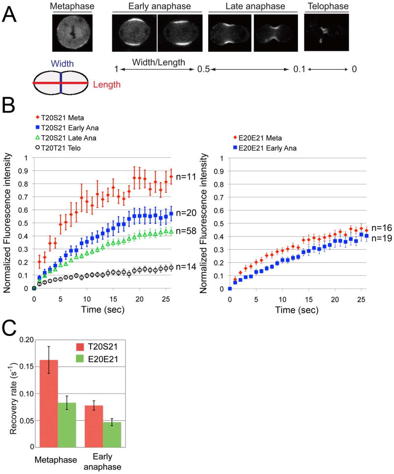
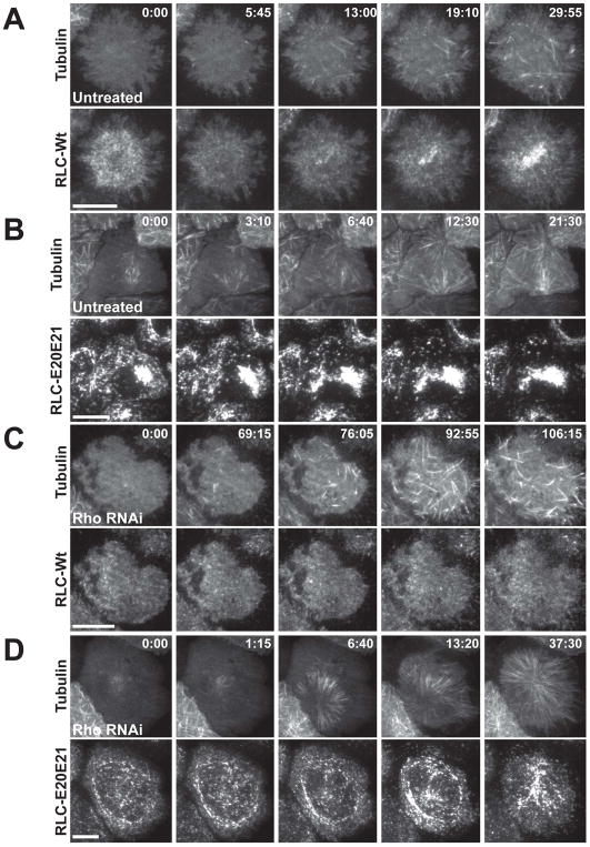
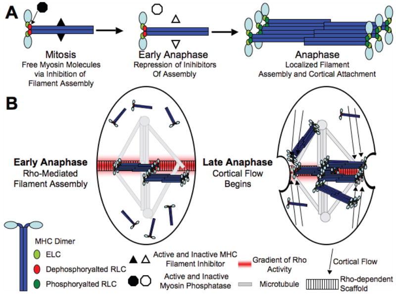
Myosin II is an essential component of the contractile ring that divides the cell during cytokinesis. Previous work showed that regulatory light chain (RLC) phosphorylation is required for localization of myosin at the cellular equator. However, the molecular mechanisms that concentrate myosin at the site of furrow formation remain unclear. By analyzing the spatiotemporal dynamics of mutant myosin subunits in Drosophila S2 cells, we show that myosin accumulates at the equator through stabilization of interactions between the cortex and myosin filaments and that the motor domain is dispensable for localization. Filament stabilization is tightly controlled by RLC phosphorylation. However, we show that regulatory mechanisms other than RLC phosphorylation contribute to myosin accumulation at three different stages: (1) turnover of thick filaments throughout the cell cycle, (2) myosin heavy chain-based control of myosin assembly at the metaphase-anaphase transition, and (3) redistribution and/or activation of myosin binding sites at the equator during anaphase. Surprisingly, the third event can occur to a degree in a Rho-independent fashion, gathering preassembled filaments to the equatorial zone via cortical flow. We conclude that multiple regulatory pathways cooperate to control myosin localization during mitosis and cytokinesis to ensure that this essential biological process is as robust as possible.
Copyright 2010 Elsevier Ltd. All rights reserved.
Figures
Similar articles
-
Myosin II recruitment during cytokinesis independent of centralspindlin-mediated phosphorylation.J Biol Chem. 2009 Oct 2;284(40):27377-83. doi: 10.1074/jbc.M109.028316. Epub 2009 Aug 6. J Biol Chem. 2009. PMID: 19661065 Free PMC article.
-
Rho kinase's role in myosin recruitment to the equatorial cortex of mitotic Drosophila S2 cells is for myosin regulatory light chain phosphorylation.PLoS One. 2006 Dec 27;1(1):e131. doi: 10.1371/journal.pone.0000131. PLoS One. 2006. PMID: 17205135 Free PMC article.
-
Multiple mechanisms for accumulation of myosin II filaments at the equator during cytokinesis.Traffic. 2008 Dec;9(12):2089-99. doi: 10.1111/j.1600-0854.2008.00837.x. Epub 2008 Sep 25. Traffic. 2008. PMID: 18939956
-
Myosin light chain kinases and phosphatase in mitosis and cytokinesis.Arch Biochem Biophys. 2011 Jun 15;510(2):76-82. doi: 10.1016/j.abb.2011.03.002. Epub 2011 Mar 21. Arch Biochem Biophys. 2011. PMID: 21396909 Free PMC article. Review.
-
Signaling pathways regulating Dictyostelium myosin II.J Muscle Res Cell Motil. 2002;23(7-8):703-18. doi: 10.1023/a:1024467426244. J Muscle Res Cell Motil. 2002. PMID: 12952069 Review.
Cited by
-
Equatorial Non-muscle Myosin II and Plastin Cooperate to Align and Compact F-actin Bundles in the Cytokinetic Ring.Front Cell Dev Biol. 2020 Sep 25;8:573393. doi: 10.3389/fcell.2020.573393. eCollection 2020. Front Cell Dev Biol. 2020. PMID: 33102479 Free PMC article.
-
Network Contractility During Cytokinesis-from Molecular to Global Views.Biomolecules. 2019 May 18;9(5):194. doi: 10.3390/biom9050194. Biomolecules. 2019. PMID: 31109067 Free PMC article. Review.
-
Myosin concentration underlies cell size-dependent scalability of actomyosin ring constriction.J Cell Biol. 2011 Nov 28;195(5):799-813. doi: 10.1083/jcb.201101055. J Cell Biol. 2011. PMID: 22123864 Free PMC article.
-
Cell shape regulation through mechanosensory feedback control.J R Soc Interface. 2015 Aug 6;12(109):20150512. doi: 10.1098/rsif.2015.0512. J R Soc Interface. 2015. PMID: 26224568 Free PMC article.
-
A novel tropomyosin isoform functions at the mitotic spindle and Golgi in Drosophila.Mol Biol Cell. 2015 Jul 1;26(13):2491-504. doi: 10.1091/mbc.E14-12-1619. Epub 2015 May 13. Mol Biol Cell. 2015. PMID: 25971803 Free PMC article.
References
Publication types
MeSH terms
Substances
Grants and funding
LinkOut - more resources
Full Text Sources
Molecular Biology Databases
