. 2010 Jun 9;18(6):700-9.
doi: 10.1016/j.str.2010.03.010.

Evolution in action: N and C termini of subunits in related T = 4 viruses exchange roles as molecular switches

Affiliations

Evolution in action: N and C termini of subunits in related T = 4 viruses exchange roles as molecular switches

Jeffrey A Speir et al. Structure. .

Abstract

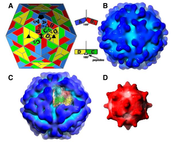
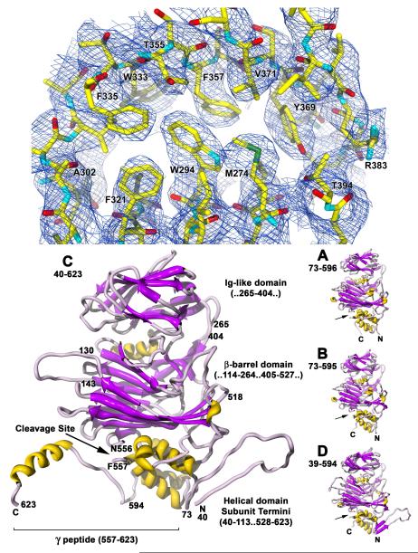
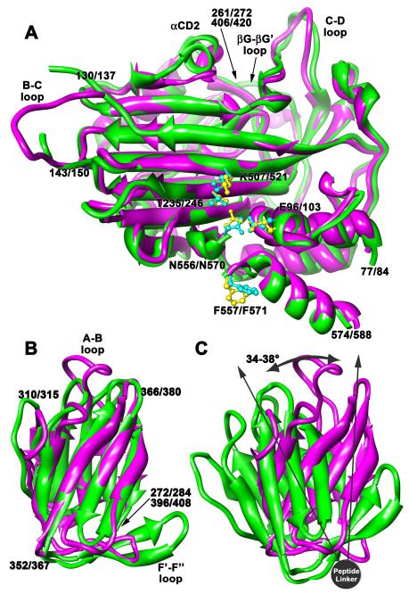
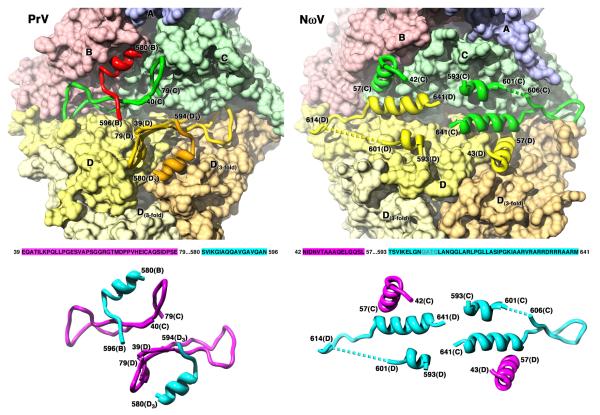
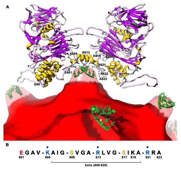
The T = 4 tetravirus and T = 3 nodavirus capsid proteins undergo closely similar autoproteolysis to produce the N-terminal beta and C-terminal, lipophilic gamma polypeptides. The gamma peptides and the N termini of beta also act as molecular switches that determine their quasi equivalent capsid structures. The crystal structure of Providence virus (PrV), only the second of a tetravirus (the first was NomegaV), reveals conserved folds and cleavage sites, but the protein termini have completely different structures and the opposite functions of those in NomegaV. N termini of beta form the molecular switch in PrV, whereas gamma peptides play this role in NomegaV. PrV gamma peptides instead interact with packaged RNA at the particle two-folds by using a repeating sequence pattern found in only four other RNA- or membrane-binding proteins. The disposition of peptide termini in PrV is closely related to those in nodaviruses, suggesting that PrV may be closer to the primordial T = 4 particle than NomegaV.


Figures

Figure 1
Figure 1
Electron cyro-microscopy reconstruction of Providence virus. (A) A T=4 quasi-symmetry model of the tetravirus capsids. Positions of icosahedral and quasi-icosahedral rotations axes are shown as filled and unfilled geometric symbols, respectively (oval = twofold; triangle = threefold; pentagon = fivefold; hexagon = sixfold). The A, B, and C polygons related by a quasi 3-fold, and the D polygon related to C by a quasi 2-fold, define the icosahedral asymmetric unit (ABCD). Each of the polygons represent identical protein subunits but occupy slightly different geometrical (chemical) environments. Polygons with subscripts are related to those without by the icosahedral symmetry of the subscript (i.e. A to A5 by 5-fold rotation). Unlike T=3 capsids, there is no icosahedral 2-fold dimer. Instead, the icosahedral 2-folds are coincident with quasi 6-fold arrangements of B, C, and D subunits (3 sets of dimers). Looking at the arrangements of ABC and DDD subunit triangles clarifies tetravirus capsid architecture. In a clear break from quasi equivalence, ABC triangles form a bent interface with each other and ABC-DDD triangles form a flat interface due to the insertion of subunit polypeptides at the interface. (B) Surface representation of the NωV reconstruction at approximately 21 Å resolution and in the same orientation as (A). Darker blue areas are at a greater radius from the particle center. The subunit Ig-like domains form large, contiguous triangular facets with curved edges around the icosahedral 3-fold axes. (C) Surface representation of PrV at approximately 28 Å resolution (same coloring and orientation as A and B). The subunits of one icosahedral asymmetric unit are shown as ribbons through their corresponding transparent surface. The most distinctive difference from NωV is that the triangular facets now have nearly straight edges and 3-fold related pits (characteristic of betatetraviruses) due to a change in the orientation of the Ig-like domains. (D) Surface representation of the PrV RNA core (same orientation as A, B, C) after removing density corresponding to the crystal structure protein coordinates. Darker red areas are at a greater radius from the particle center. Large bulges of density extend from the core at each icosahedral 2-fold axis and make contact with the capsid protein shell.
Figure 2
Figure 2
The crystal structure of Providence virus. (Top) Electron density from the C subunit Ig-like domain core contoured at approximately 1.5σ. The beta strands and side chains are clearly delineated in the 30-fold averaged map. (Bottom) Ribbon representation of all four quasi equivalent subunits (top – capsid exterior, bottom – capsid interior). The C subunit is enlarged to show the different domains and their residue ranges (labeled to the right), the cleavage site (labeled with arrows in all 4 subunits), and its extended gamma peptide (underlined). The residue range fitted is shown underneath each subunit letter. Note that subunits A, B, and D do not have an extended gamma peptide as see in C, but the D subunit has an extended N-terminus with an almost identical structure to that of C.
Figure 3
Figure 3
Tetravirus structure comparison. (A) Superposition of the beta barrel and core helical domains of the PrV (magenta) and NωV (green) C subunits viewed tangential to the protein shell. Residues 77-261 and 406-574 from PrV were aligned with residues 84-272 and 420-588 of NωV. Labels list the PrV residue number first, followed by a slash, then the NωV residue number. The r.m.s.d is 1.5Å and the sequence identity is 47% for 350/356 aligned residues. The autoproteolysis sites have identical key residues, shown in blue for Prv and yellow for NωV, and closely similar structures. The Ig-like domains would be attached at the positions designated by the long arrow at the top. (B) Superposition of the Ig-like domains from the C subunits (same coloring and labeling as above). Residues 272-396 of PrV were aligned with residues 284-408 of NωV. More variation occurs in these domains, mainly in the turns between beta strands, resulting in an r.m.s.d of 2.4Å. The sequence identity is only 15% for 113/125 aligned residues. (C) The orientations of the C-subunit Ig-like domains when the beta barrels are aligned as in (A). The domains have strikingly different rotations about the peptide linker that range between 34-38° depending on the subunit.
Figure 4
Figure 4
Opposite roles for PrV polypeptides forming the molecular switch. (Top) Inside surface representation of the flat contacts. Subunits are shown colored as in Fig. 1A, except that the three D subunits are differentiated using shades of yellow to gold. The ordered polypeptides that form the switch are shown as ribbons without their corresponding surfaces. In PrV, extended N-termini from C (40-79) and D (39-79) fill the groove, and are supported by the last ordered residues of the C-terminal gamma peptides from quasi or icosahedral 3-fold related subunits (B580-B596 and D580-D594, respectively). In NωV, the same roles for the polypeptides are reversed. Extended C-terminal gamma peptides from the C and D subunits (593-641) fill the groove and are supported by the first ordered residues of the quasi 2-fold related subunit: D43-57 supports the C subunit gamma peptide, and C42-57 supports the D subunit gamma peptide. (Bottom) Diagram showing just the molecular switches from PrV and NωV. The relevant capsid protein sequence of each section is shown and color coded as the N-terminal peptides (magenta) or the C-terminal peptides (blue). The structure of each is shown with the same color coding, demonstrating the swap of N-termini for C-termini between PrV and NωV for the same roles in forming the flat contacts.
Figure 5
Figure 5
Protein-RNA contacts between the C subunit gamma peptides and packaged genome in PrV. (A) The gamma peptide structures create a concave volume 28Å in diameter containing 12 charged residues (10 basic, 2 acidic) that is filled with RNA. One pair of icosahedral 2-fold related C subunits (ribbons) are shown above the RNA core (red) viewed tangential to the capsid shell. The RNA protruding at the 2-folds (Fig. 1D) has been made transparent to show the small section of partially ordered RNA (green) observed in the crystal structure. Side chains with key interactions are shown on the anti-parallel helices sitting over the RNA (only those on the front helix are numbered). (B) Sequence of the gamma peptide at the site of RNA interactions. Acidic residues are red, basic are blue, and other hydrophilics are gold. Residues with asterisks directly contact the ordered RNA. A pattern of a single hydrophilic residue followed by 3 hydrophobic residues repeats 5 times, which structurally forms a hydrophobic zipper between the anti-parallel helices with the hydrophilic residues oriented downward toward the RNA. Sequence searches using this pattern revealed some similarities to other RNA and membrane binding proteins.

Similar articles

Cited by

References

    1. Altschul SF, Madden TL, Schaffer AA, Zhang J, Zhang Z, Miller W, Lipman DJ. Gapped BLAST and PSI-BLAST: a new generation of protein database search programs. Nucleic Acids Res. 1997;25:3389–3402. - PMC - PubMed
    1. Bahadur RP, Zacharias M, Janin J. Dissecting protein-RNA recognition sites. Nucleic Acids Res. 2008;36:2705–2716. - PMC - PubMed
    1. Baker ML, Jiang W, Rixon FJ, Chiu W. Common ancestry of herpesviruses and tailed DNA bacteriophages. J Virol. 2005;79:14967–14970. - PMC - PubMed
    1. Banerjee M, Khayat R, Walukiewicz HE, Odegard AL, Schneemann A, Johnson JE. Dissecting the functional domains of a nonenveloped virus membrane penetration peptide. J Virol. 2009;83:6929–6933. - PMC - PubMed
    1. Bothner B, Dong XF, Bibbs L, Johnson JE, Siuzdak G. Evidence of viral capsid dynamics using limited proteolysis and mass spectrometry. J Biol Chem. 1998;273:673–676. - PubMed

Publication types

Substances

LinkOut - more resources