Review
. 2010 Jul;65(7):567-81.
doi: 10.1016/j.crad.2010.02.013.

Molecular ultrasound imaging: current status and future directions

Affiliations
Review

Molecular ultrasound imaging: current status and future directions

N Deshpande et al. Clin Radiol. 2010 Jul.

Abstract

Targeted contrast-enhanced ultrasound (molecular ultrasound) is an emerging imaging strategy that combines ultrasound technology with novel molecularly-targeted ultrasound contrast agents for assessing biological processes at the molecular level. Molecular ultrasound contrast agents are nano- or micro-sized particles that are targeted to specific molecular markers by adding high-affinity binding ligands onto the surface of the particles. Following intravenous administration, these targeted ultrasound contrast agents accumulate at tissue sites overexpressing specific molecular markers, thereby enhancing the ultrasound imaging signal. High spatial and temporal resolution, real-time imaging, non-invasiveness, relatively low costs, lack of ionising irradiation and wide availability of ultrasound systems are advantages compared to other molecular imaging modalities. In this article we review current concepts and future directions of molecular ultrasound imaging, including different classes of molecular ultrasound contrast agents, ongoing technical developments of pre-clinical and clinical ultrasound systems, the potential of molecular ultrasound for imaging different diseases at the molecular level, and the translation of molecular ultrasound into the clinic.


Figures

Figure 1
Figure 1
Different types of ultrasound contrast agents. a) Microbubbles are gas-liquid emulsions with a polyethylene gycol (PEG) polymer on the surface to prevent aggregation. Microbubbles are highly echogenic and the most commonly used contrast agent for molecular ultrasound imaging. b) Perfluorocarbon emulsion (PFC) nanodroplets are liquid-liquid emulsions that can be vaporized into echogenic gas-bubbles following administration of acoustic energy. c) Liposomes are phospholipid bilayers that can enclose air pockets for ultrasound imaging. d) Nanobubbles are gas-liquid emulsions that can fuse into echogenic microbubbles at the target site. e) Solid nanoparticles are solid amorphous substances with gas entrapped in their pores or fissures increasing echogenicity.
Figure 2
Figure 2
Schematic representation of the use of PFC nanodroplets as ultrasound contrast agents. Due to their small size, PFC nanodroplets extravasate into the tumor interstitium. Following administration of acoustic energy, the liquid perfluorocarbon core can be vaporized and transferred into gaseous phase, thereby increasing echogenicity at the site of PFC nanodroplet accumulation.
Figure 3
Figure 3
Schematic representation of the use of nanobubbles as ultrasound contrast agents. Tumors are characterized by defective vasculature with large gaps between the endothelial cells, allowing extravasation of non-targeted nanobubbles and passive accumulation in tumor interstitium. In the tumor interstitium, these nanobubbles can coalesce to form echogenic microbubbles, detectable by ultrasound imaging (modified from Rapport et al, J Natl Cancer Inst 2007).
Figure 4
Figure 4
a) Schematic representation of targeted microbubbles (MB) attached to receptors expressed on tumor endothelial cells after intravenous administration. Microbubbles remain predominantly in the vasculature due to their size (several micrometers) and thus adhere only to the tumor endothelial cells and not to tumor cells. Some microbubbles do not attach to receptors and freely float. After high-power destructive pulse, adherent microbubbles are destroyed and freely circulating microbubbles replenish from outside imaging plane after several seconds. b) Graphical summary of the approach for quantification of imaging signal from attached microbubbles. Adapted from Willmann et al, Radiology Vol 246, number 2, 2008 .
Figure 5
Figure 5
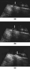
Molecular ultrasound images of a subcutaneous human ovarian adenocarcinoma xenograft tumor (arrows) in a nude mouse after intravenous administration of singly-targeted microbubbles, targeted at vascular endothelial growth factor receptor type 2 (a) or αvβ3 integrin (b), and dual-targeted microbubbles (c). Molecular ultrasound imaging signal is shown as green overlay on B-mode images. Note that in vivo imaging signal is substantially higher after administration of dual-targeted microbubbles compared to either of the singly-targeted microbubbles. Reprinted with permission from Willmann et al, Radiology Vol 248, number 3, 2008 .
Figure 6
Figure 6
Dual-targeted microbubble can be designed that mimic the behavior of leukocytes in vivo. a) Circulating leukocytes in the blood invade the site of inflammation through a process of several steps: 1) Capturing and 2) rolling of leukocytes along the vasculature mediated through transient interactions between selectin proteins (e.g., P-selectin) and their ligands (e.g., P-selectin glycoprotein ligand-1 - PSGL-1); 3) Firm adhesion of leukocytes to endothelial cells by high affinity interaction between cell adhesion molecules (e.g., VCAM1) and integrins (e.g., Very Late Antigen-4- VLA-4); and 4) Extravasation (modified from Kunkel and Butcher 2003, Nature Reviews Immunology 3, 822-829 ). b) Dual-targeted microbubble targeted against both P-selectin and VCAM1, simulating vascular attachment of a leukocyte at sites of inflammation by first interacting with P-selectin and then firmly attaching via VCAM1.
Figure 7
Figure 7
Inflammation imaging in inflammatory bowel disease using contrast enhanced transabdominal ultrasound and microbubbles targeted at MAdCAM-1 (Mucosal Addressin Cellular Adhesion Molecule). A) anatomical image of excised shows terminal ileum (TI) and enlarged draining mesenteric lymph node (MLN). B) Corresponding in vivo color-coded molecular ultrasound image shows inflammation in TI and MLN. Reprinted with permission from Bachmann et al, Gastroenterology, Vol 130: 8-16, 2006 .
Figure 8
Figure 8
Molecular ultrasound images of thrombi in femoral vein in the hind limb of dogs. A) at baseline without any contrast agent b) after the administration of non targeted microbubbles (SonoVue) c) after the administration of microbubbles targeted against GP (Platelet Glycoprotein) IIb/IIa receptor. Note better visualization of intravascular thrombus with targeted microbubbles. Reprinted with permission from Wang et al, Acad Radiol, Vol 13: 428-433, 2006 .
Figure 9
Figure 9
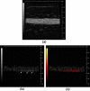
(a) High-frequency (30 MHz) B-Mode image of a biotinylated gelatin vessel phantom which is perfused with avidinated microbubbles. (b) Nonlinear subharmonic (15 MHz) image of the avidinated microbubbles inside the phantom, without interframe filtering signal processing (microbubbles bound to the vessel wall through the avidin-biotin complex are highlighted with white arrows). (c) Subharmonic image of avidinated microbubbles inside the phantom, using interframe filtering to isolate signal from bound microbubbles in the presence of flowing ones (bound microbubbles highlighted with red overlay) Reprinted with permission from Needles et al, Ultrasound Med Biol, 35(9): 1564-1573, 2009.

Similar articles

Cited by

References

    1. Lanza GM, Wickline SA. Targeted ultrasonic contrast agents for molecular imaging and therapy. Curr Probl Cardiol. 2003;28(12):625. - PubMed
    1. Klibanov AL. Molecular imaging with targeted ultrasound contrast microbubbles. Ernst Schering Res Found Workshop. 2005;(49):171. - PubMed
    1. McCulloch M, Gresser C, Moos S, et al. Ultrasound contrast physics: A series on contrast echocardiography, article 3. J Am Soc Echocardiogr. 2000;13(10):959. - PubMed
    1. Klibanov A, Gu H, Wojdyla JK, et al. Attachemnet of ligands to gas filled microbubbles via PEG spacer and lipid residues anchored at the interface. Boston: 1999. p. 124.
    1. Walday P, Tolleshaug H, Gjoen T, et al. Biodistributions of air-filled albumin microspheres in rats and pigs. Biochem J. 1994;299(Pt 2):437. - PMC - PubMed

Publication types

MeSH terms