

Glucocorticoid-induced apoptosis of healthy and malignant lymphocytes
- PMID: 20541659
- PMCID: PMC4770454
- DOI: 10.1016/S0079-6123(10)82001-1
Glucocorticoid-induced apoptosis of healthy and malignant lymphocytes
Abstract
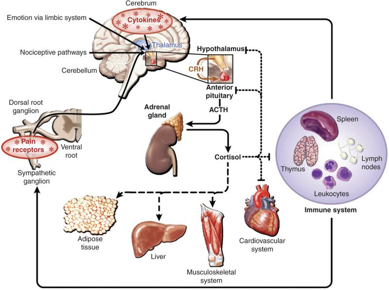
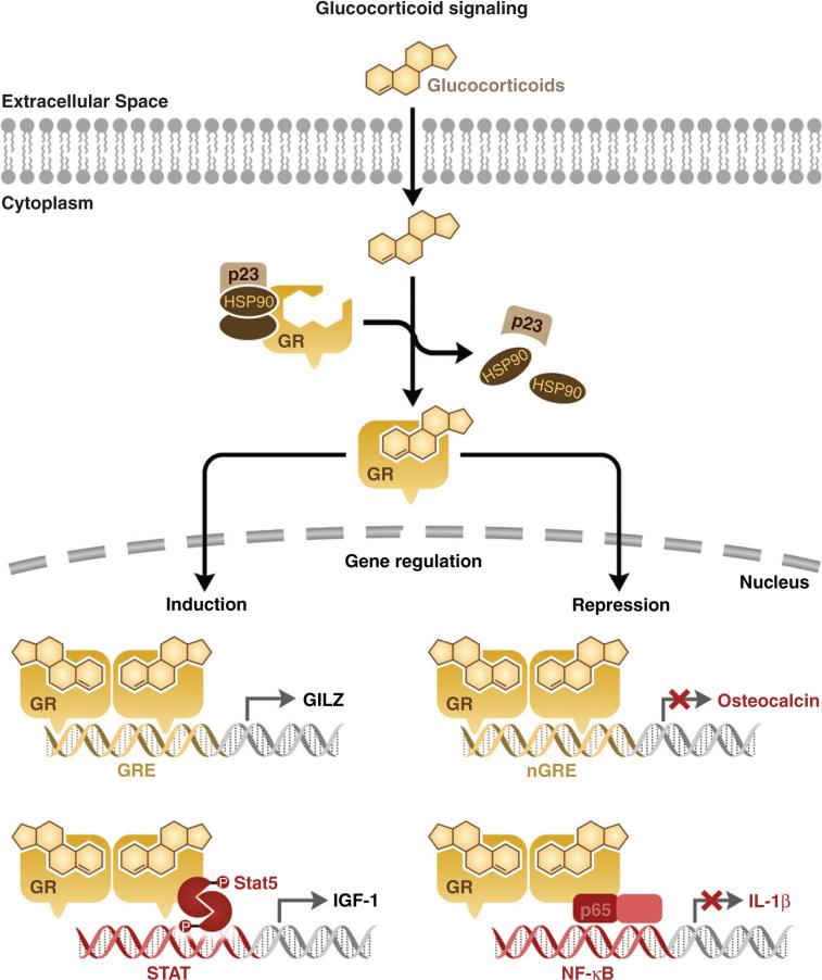
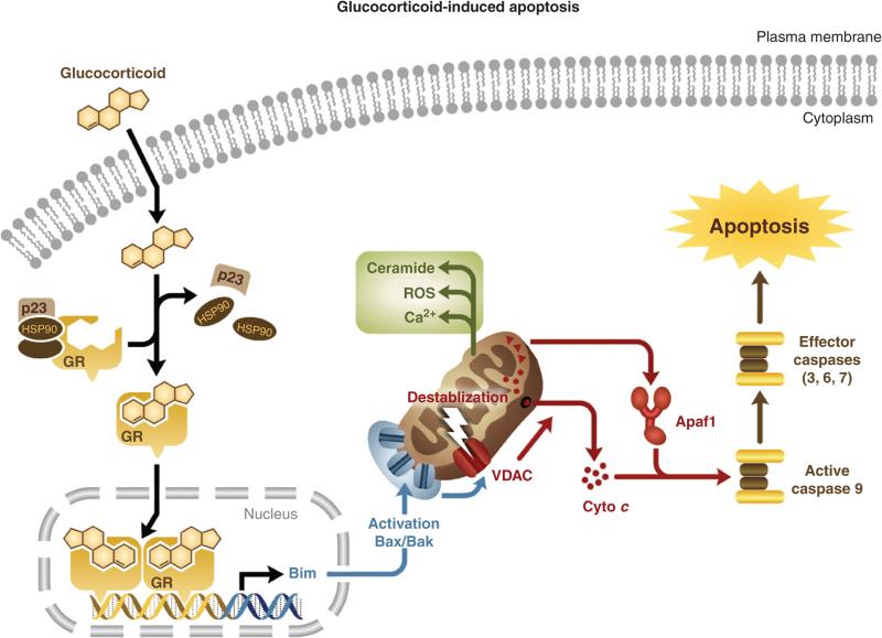
Glucocorticoids exert a wide range of physiological effects, including the induction of apoptosis in lymphocytes. The progression of glucocorticoid-induced apoptosis is a multi-component process requiring contributions from both genomic and cytoplasmic signaling events. There is significant evidence indicating that the transactivation activity of the glucocorticoid receptor is required for the initiation of glucocorticoid-induced apoptosis. However, the rapid cytoplasmic effects of glucocorticoids may also contribute to the glucocorticoid-induced apoptosis-signaling pathway. Endogenous glucocorticoids shape the T-cell repertoire through both the induction of apoptosis by neglect during thymocyte maturation and the antagonism of T-cell receptor (TCR)-induced apoptosis during positive selection. Owing to their ability to induce apoptosis in lymphocytes, synthetic glucocorticoids are widely used in the treatment of haematological malignancies. Glucocorticoid chemotherapy is limited, however, by the emergence of glucocorticoid resistance. The development of novel therapies designed to overcome glucocorticoid resistance will dramatically improve the efficacy of glucocorticoid therapy in the treatment of haematological malignancies.
Copyright 2010 Elsevier B.V. All rights reserved.
Figures
Similar articles
-
Glucocorticoid-induced apoptosis in lymphocytes.Biochem Biophys Res Commun. 2000 Dec 20;279(2):307-12. doi: 10.1006/bbrc.2000.3922. Biochem Biophys Res Commun. 2000. PMID: 11118283 Review.
-
Mechanisms of glucocorticoid-induced apoptosis in hematologic malignancies: updates.Curr Opin Oncol. 2004 Nov;16(6):553-63. doi: 10.1097/01.cco.0000142072.22226.09. Curr Opin Oncol. 2004. PMID: 15627017 Review.
-
[Induction of apoptosis in lymphocytes by glucocorticoids: between physiology and pharmacology].C R Seances Soc Biol Fil. 1998;192(6):1051-63. C R Seances Soc Biol Fil. 1998. PMID: 10101602 Review. French.
-
Stepping stones in the path of glucocorticoid-driven apoptosis of lymphoid cells.Acta Biochim Biophys Sin (Shanghai). 2008 Jul;40(7):595-600. doi: 10.1111/j.1745-7270.2008.00433.x. Acta Biochim Biophys Sin (Shanghai). 2008. PMID: 18604450 Review.
-
Molecular mechanisms of glucocorticoids in the control of inflammation and lymphocyte apoptosis.Crit Rev Clin Lab Sci. 2005;42(1):71-104. doi: 10.1080/10408360590888983. Crit Rev Clin Lab Sci. 2005. PMID: 15697171 Review.
Cited by
-
The Pro-inflammatory Effects of Glucocorticoids in the Brain.Front Endocrinol (Lausanne). 2016 Jun 28;7:78. doi: 10.3389/fendo.2016.00078. eCollection 2016. Front Endocrinol (Lausanne). 2016. PMID: 27445981 Free PMC article. Review.
-
miR-221/222-Mediated Inhibition of Autophagy Promotes Dexamethasone Resistance in Multiple Myeloma.Mol Ther. 2019 Mar 6;27(3):559-570. doi: 10.1016/j.ymthe.2019.01.012. Epub 2019 Jan 24. Mol Ther. 2019. PMID: 30765325 Free PMC article.
-
Serum neurofilament light chains in progressive multiple sclerosis patients treated with repeated cycles of high-dose intravenous steroids.Free Neuropathol. 2023 Oct 5;4:15. doi: 10.17879/freeneuropathology-2023-5049. eCollection 2023 Jan. Free Neuropathol. 2023. PMID: 37859628 Free PMC article.
-
Relapse-associated AURKB blunts the glucocorticoid sensitivity of B cell acute lymphoblastic leukemia.Proc Natl Acad Sci U S A. 2019 Feb 19;116(8):3052-3061. doi: 10.1073/pnas.1816254116. Epub 2019 Feb 7. Proc Natl Acad Sci U S A. 2019. PMID: 30733284 Free PMC article.
-
Oncogenic deubiquitination controls tyrosine kinase signaling and therapy response in acute lymphoblastic leukemia.Sci Adv. 2022 Dec 9;8(49):eabq8437. doi: 10.1126/sciadv.abq8437. Epub 2022 Dec 9. Sci Adv. 2022. PMID: 36490346 Free PMC article.
References
-
- Abrams MT, Robertson NM, Yoon K, Wickstrom E. Inhibition of glucocorticoid-induced apoptosis by targeting the major splice variants of BIM mRNA with small interfering RNA and short hairpin RNA. The Journal of Biological Chemistry. 2004;279:55809–55817. - PubMed
-
- Adcock IM. Glucocorticoid-regulated transcription factors. Pulmonary Pharmacology & Therapeutics. 2001;14:211–219. - PubMed
-
- Alksnis M, Barkhem T, Stromstedt PE, Ahola H, Kutoh E, Gustafsson JA, et al. High level expression of functional full length and truncated glucocorticoid receptor in Chinese hamster ovary cells. Demonstration of ligand-induced down-regulation of expressed receptor mRNA and protein. The Journal of Biological Chemistry. 1991;266:10078–10085. - PubMed
-
- Ashkenazi A, Dixit VM. Death receptors: Signaling and modulation. Science. 1998;281:1305–1308. - PubMed
-
- Ashwell JD, Lu FW, Vacchio MS. Glucocorticoids in T cell development and function . Annual Review of Immunology. 2000;18:309–345. - PubMed
MeSH terms
Substances
Grants and funding
LinkOut - more resources
Full Text Sources
Other Literature Sources
Medical
