. 2010 Jun 15;17(6):560-73.
doi: 10.1016/j.ccr.2010.04.023.

NSAID sulindac and its analog bind RXRalpha and inhibit RXRalpha-dependent AKT signaling

Affiliations

NSAID sulindac and its analog bind RXRalpha and inhibit RXRalpha-dependent AKT signaling

Hu Zhou et al. Cancer Cell. .

Abstract

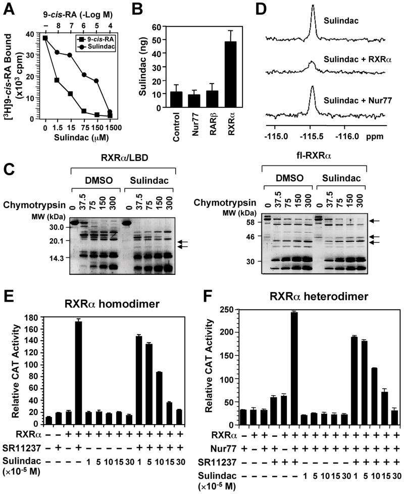
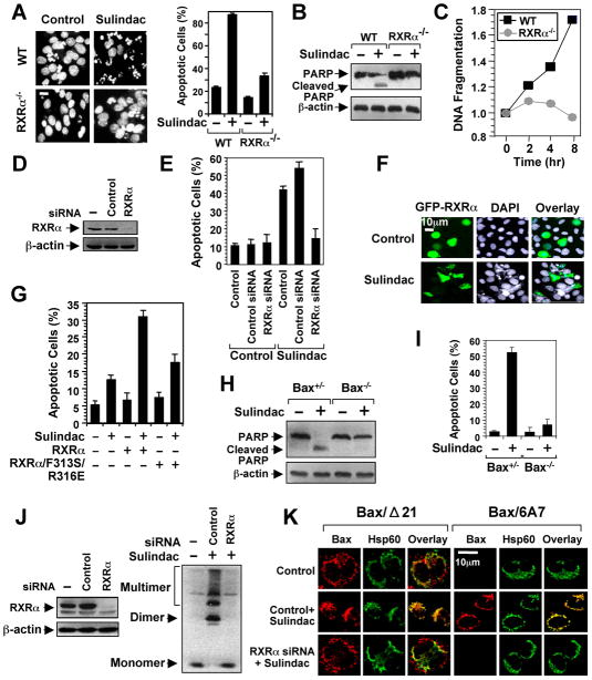
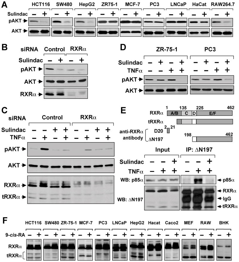
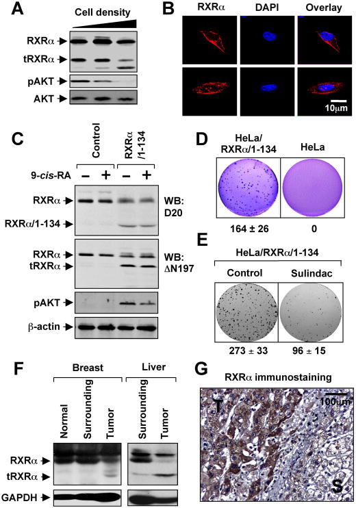
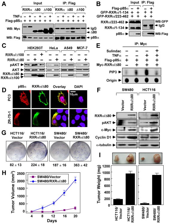
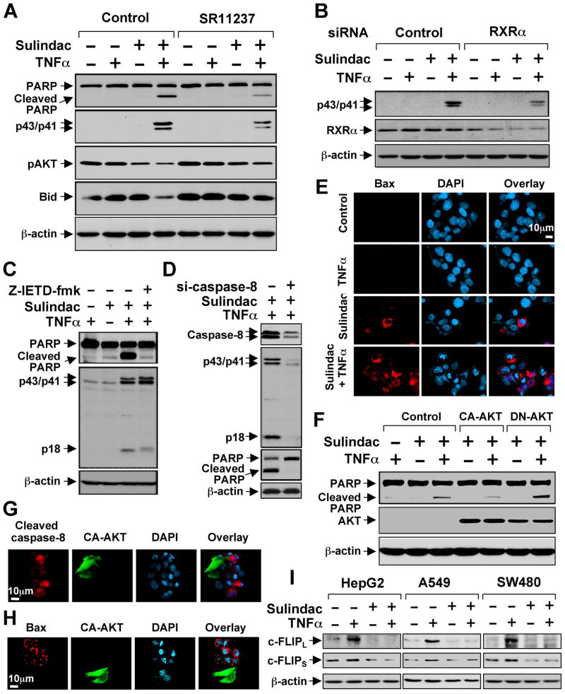
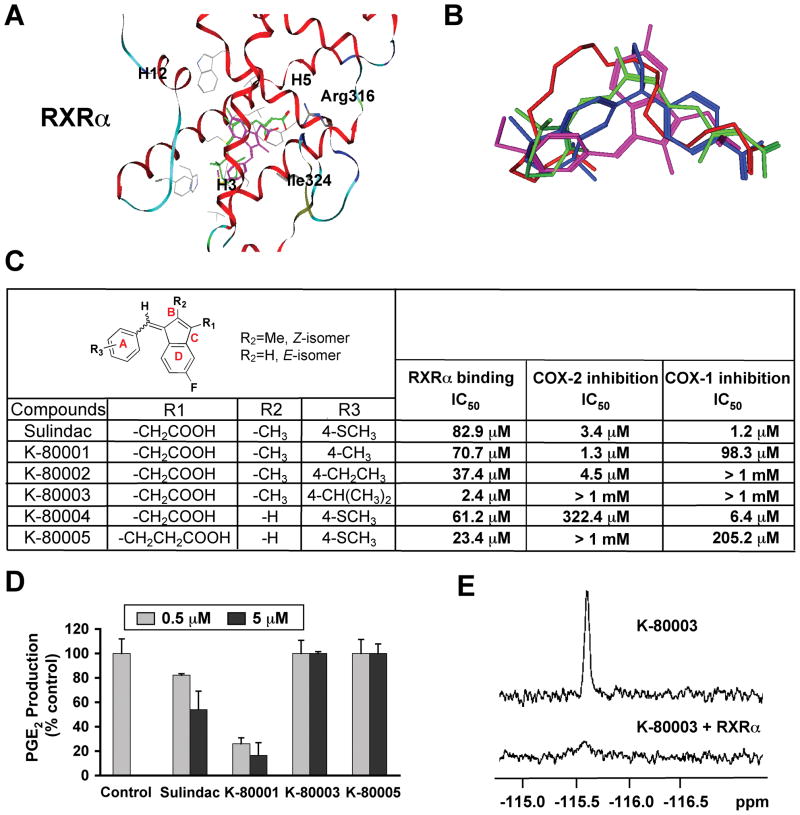
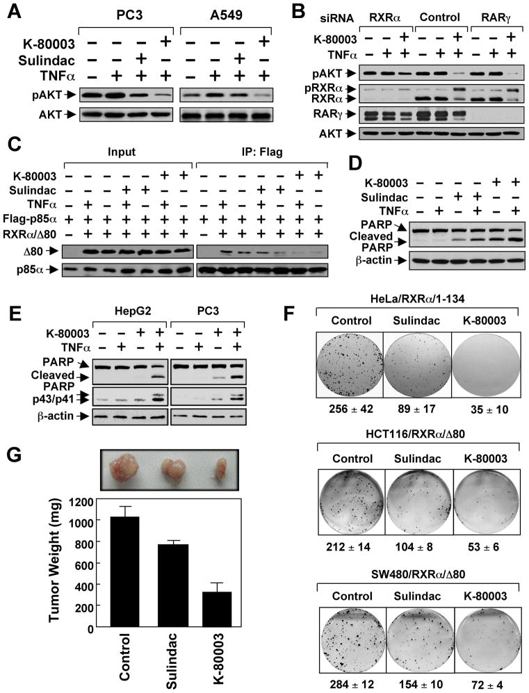
Nonsteroidal anti-inflammatory drugs (NSAIDs) exert their anticancer effects through cyclooxygenase-2 (COX-2)-dependent and independent mechanisms. Here, we report that Sulindac, an NSAID, induces apoptosis by binding to retinoid X receptor-alpha (RXRalpha). We identified an N-terminally truncated RXRalpha (tRXRalpha) in several cancer cell lines and primary tumors, which interacted with the p85alpha subunit of phosphatidylinositol-3-OH kinase (PI3K). Tumor necrosis factor-alpha (TNFalpha) promoted tRXRalpha interaction with the p85alpha, activating PI3K/AKT signaling. When combined with TNFalpha, Sulindac inhibited TNFalpha-induced tRXRalpha/p85alpha interaction, leading to activation of the death receptor-mediated apoptotic pathway. We designed and synthesized a Sulindac analog K-80003, which has increased affinity to RXRalpha but lacks COX inhibitory activity. K-80003 displayed enhanced efficacy in inhibiting tRXRalpha-dependent AKT activation and tRXRalpha tumor growth in animals.


Figures

Figure 1
Figure 1. Sulindac Binds to RXRα
(A) Sulindac binding to RXRα in vitro. RXRα LBD protein was incubated with [3H]9-cis-RA in the presence or absence of Sulindac or unlabeled 9-cis-RA. Bound [3H]9-cis-RA was quantitated by liquid scintillation counting. (B) Sulindac binding to RXRα in cells. HEK293 cells stably expressing RXRα, Nur77 or RARβ were treated with 100 μM sulindac for 3 hr. The same amount of purified RXRα, Nur77, or RARβ protein was subjected to HPLC analysis for the presence of Sulindac. (C) Altered sensitivity of RXRα LBD or GST-RXRα to chymotrypsin (μg/ml) by Sulindac (100 μM). (D) Comparison of 19F NMR spectra of Sulindac (100 μM) in the absence and presence of 10 μM RXRα LBD or Nur77 protein. (E, F) Sulindac inhibits transactivation of RXRα homodimers and heterodimers. (TREpal)2-tk-CAT (Zhang et al., 1992a) (E) or βRARE-tk-CAT (Zhang et al., 1992b) (F), Nur77 and/or RXRα were transiently transfected into CV-1 cells. Cells were treated with or without SR11237 (10−6 M) in the presence or absence of Sulindac. CAT activity was determined. One of three to five similar experiments is shown. Error bars represent SEM. See also Figure S1.
Figure 2
Figure 2. Sulindac Induces RXRα-dependent Apoptosis and Bax Activation
(A–C) The apoptotic effects of Sulindac in F9 or F9 cells lacking RXRα (F9 RXRα−/−). Cells treated with Sulindac (75 μM) for 24 hr were analyzed by DAPI staining (A), PARP cleavage (B), and DNA fragmentation (C). Scale bar: 10 μm. (D,E) RXRα siRNA inhibits apoptosis induction by Sulindac. H460 lung cancer cells transfected with control or RXRα siRNA were treated with Sulindac (75 μM) for 24 hr and analyzed by DAPI staining for apoptosis. (F) Transfection of RXRα enhances the apoptotic effect of Sulindac. CV-1 cells transfected with GFP-RXRα were treated with Sulindac (75 μM) for 24 hr and analyzed by DAPI staining. GFP-RXRα-transfected cells underwent extensive nuclear fragmentation and condensation. (G) Disruption of the RXRα LBP impairs the apoptotic effect of Sulindac. CV-1 cells transfected with GFP-RXRα or GFP-RXRα/F313S/R316E were treated with Sulindac (75 μM) for 24 hr and analyzed by DAPI staining. Apoptosis scored in receptor-transfected cells. See also Figure S2. (H,I) Role of Bax in apoptosis induction by Sulindac. HCT116 cells or HCT116 cells lacking Bax (Bax−/−) were treated with or without sulindac (75 μM) for 24 hr. Apoptosis determined by PARP cleavage (H) and DAPI staining (I). (J,K) RXRα siRNA inhibits sulindac-induced Bax activation. Knocking down RXRα in HCT116 cells by RXRα siRNA revealed by immunoblotting. HCT116 cells transfected with or without RXRα siRNA or control siRNA for 48 hr were treated with Sulindac for 6 hr, and analyzed for Bax oligomerization (J) and Bax conformational change and mitochondrial targeting by immunostaining/confocal microscopy using Bax/Δ21, Bax/6A7, or anti-Hsp60 antibody (K). About 60% of cells showed Bax conformational change presented. One of three to five similar experiments is shown. Error bars represent SEM.
Figure 3
Figure 3. Sulindac Inhibits TNFα-induced AKT Activation and tRXRα-p85α Interaction
(A) Inhibition of AKT activation by Sulindac. The indicated cells starved overnight and treated with Sulindac (100 μM) for 1 hr were analyzed for AKT activation by immunoblotting. (B) Inhibition of basal AKT activation by RXRα siRNA. HepG2 cells transfected with RXRα siRNA for 48 hr were treated with Sulindac (100 μM) for 1 hr. AKT activation and RXRα expression were analyzed by immunoblotting. (C) Inhibition of TNFα-induced AKT activation by Sulindac and RXRα siRNA. A549 lung cancer cells transfected with RXRα or control siRNA for 48 hr were pretreated with Sulindac (100 μM) for 1 hr before exposed to TNFα (10 ng/ml) for 30 min. (D) Synergistic inhibition of AKT activation by TNFα and Sulindac. ZR-75-1 and PC3 cells were pretreated with Sulindac for 1 hr before exposed to TNFα (10 ng/ml) for 30 min. (E) Induction of RXRα-p85α interaction by TNFα. A549 cells treated with TNFα (10 ng/ml) and/or Sulindac (100 μM) for 30 min were analyzed for RXRα-p85α interaction by co-immunoprecipitation using ΔN197 antibody. Above, schematic representation of anti-RXRα antibodies used in co-immunoprecipitation and immunoblotting assays. D20 antibody recognizes amino acids 2–21 in the N-terminal A/B domain, while ΔN197 antibody recognizes the C-terminal E/F domain. A RXRα truncated protein with about 44 kDa is also shown. (F) Expression of tRXRα in various cancer cell lines. The indicated cell lines treated with or without 9-cis-RA (10−7 M) for 30 min were analyzed by immunoblotting using the ΔN197 RXRα antibody. One of three to six similar experiments is shown. See also Figure S3.
Figure 4
Figure 4. Role of tRXRα in AKT Activation and Anchorage-Independent Cell Growth
(A) Cell density dependent production of tRXRα and AKT activation. MEFs seeded at different cell density were analyzed for RXRα expression using ΔN197 antibody and for AKT activation by immunoblotting. (B) Subcellular localization of endogenous RXRα in MEFs was visualized by confocal microscopy after immunostaining using anti-RXRα (ΔN197). Cells were also stained with DAPI to visualize the nucleus. More than 60% of cells showed the images presented. (C) Stable expression of RXRα/1–134 induces RXRα cleavage and AKT activation. HeLa or HeLa cells stably expressing RXRα/1–134 were treated with 9-cis-RA for 30 min and analyzed for AKT activation and expression of RXRα. (D) Growth of HeLa/RXRα/1–134 and HeLa cells in soft agar. (E) Sulindac inhibits clonogenic survival of HeLa/RXRα/1–134 cells. Cells grown in 6-well plates for 5 days were treated with Sulindac (25 μM) for 3 days. (F) Production of tRXRα in human tumor tissues of breast (5 out of 6) or liver (4 out of 6) compared to tumor surrounding and normal tissues. (G) Cytoplasmic localization of RXRα in liver tumor specimens immunostained by ΔN197 antibody. T, tumor tissue; S, tumor surrounding tissue. One of three to five similar experiments is shown. See also Figure S4.
Figure 5
Figure 5. Role of N-terminally Truncated RXRα in PI3K/AKT Activation by TNFα and Cancer Cell Growth
(A) HEK293T cells were transfected with Flag-p85α and RXRα, RXRα/Δ80, or RXRα/Δ100 tagged with the Myc epitope, treated with TNFα (10 ng/ml) for 30 min, and analyzed by co-immunoprecipitation using anti-Flag antibody. (B) N-terminal A/B domain of RXRα interacts with p85α. Flag-p85α was cotransfected with GFP-RXRα/1–134 and GFP-RXRα/223–462 into HEK293T cells, and analyzed for their interaction by co-immunoprecipitation using anti-Flag antibody. (C) RXRα/Δ80 is a potent AKT activator. AKT activation of the indicated cells transfected with RXRα/Δ80 or RXRα/Δ100 was determined by immunoblotting. (D) Cytoplasmic co-localization of RXRα/Δ80 and p85α. Myc-RXRα/Δ80 and p85α were cotransfected into the indicated cell lines, immunostained with anti-Myc and anti-p85α antibody, and their subcellular localization revealed by confocal microscopy. RXRα/Δ80 predominantly resided in the cytoplasm of about 80% of cells. About 15% of cells showed the images presented. (E) Activation of PI3K by RXRα/Δ80 immunoprecipitates. A549 cells transfected with Flag-p85α and Myc-RXRα/80 were treated with TNFα and/or Sulindac, immunoprecipitated with anti-Myc antibody, and subjected to in vitro PI3K assay. (F) AKT activation by stable expression of RXRα/Δ80. Cells stably transfected with GFP-RXRα/Δ80 or control GFP vector were analyzed by immunoblotting. (G) RXRα/Δ80 promotes clonogenic survival of cancer cells grown in 6-well plates for 8 days. (H,I) RXRα/Δ80 promotes cancer cell growth in nude mice (n=6) for three weeks. One of three to five similar experiments is shown. Error bars represent SEM. See also Figure S5.
Figure 6
Figure 6. Activation of TNFα-induced Extrinsic Apoptotic Pathway by Sulindac
(A) Synergistic induction of apoptosis by Sulindac/TNFα combination and its inhibition by RXRα ligand. HepG2 cells cultured in medium with 1% FBS were treated with SR11237 (1 μM) for 1 hr, then TNFα (10 ng/ml) and/or Sulindac (75 μM) for 4 hr, and analyzed by immunoblotting. (B) Inhibition of Sulindac/TNFα-induced caspases-8 cleavage by RXRα siRNA. HepG2 cells transfected with control or RXRα siRNA were treated with TNFα and/or Sulindac and analyzed by immunoblotting. (C,D) Inhibition of Sulindac/TNFα-induced PARP cleavage by caspase-8 inhibitor and siRNA. HepG2 cells transfected with control or caspase-8 siRNA or pretreated with Z-IETD-fmk (40 μM) for 1 hr were treated with TNFα and Sulindac and analyzed by immunoblotting. (E) Activation of Bax by Sulindac and TNFα. HepG2 cells treated with TNFα and/or Sulindac were immunostained with Bax/6A7 antibody. About 15% Sulindac-treated cells while about 60% Sulindac/TNFα-treated cells showed Bax staining. (F) Regulation of Sulindac/TNFα-induced PARP cleavage by AKT. PC3 cells transfected with CA-AKT or DN-AKT were treated with TNFα and/or Sulindac, and analyzed by immunoblotting. (G,H) CA-AKT inhibits activation of caspase-8 (G) and Bax (H) by Sulindac and TNFα. HepG2 cells transfected with CA-AKT were treated with TNFα and Sulindac, and immunostained with anti-cleaved caspase-8 or Bax/6A7 antibody. About 80% nontransfected and 15% CA-AKT-transfected cells showed caspase-8 staining. About 60% nontransfected and about 13% CA-AKT-transfected cells exhibited Bax staining. (I) Regulation of c-FLIP expression by TNFα and Sulindac. Cells treated with TNFα and/or Sulindac for 6 hr were analyzed by immunoblotting. One of three to five similar experiments is shown. See also Figure S6.
Figure 7
Figure 7. Design, Synthesis and Evaluation of RXRα-selective Sulindac Analogs
(A) Docking of sulindac sulfide (magenta) to the LBP of RXRα in reference to 9-cis-RA (green). Side chains within 4Å of the ligands are displayed in grey. (B) Comparison of orientation and position of docked sulindac sulfide (magenta) to the crystal structures of 9-cis-RA (green), DHA (red) and BMS649 (blue). (C) RXRα binding and inhibition of COX-1 and COX-2 activities by Sulindac analogs. RXRα binding was measured by competition ligand-binding assays. COX inhibition assays used Cayman’s COX Fluorescent Activity Assay Kits. (D) Inhibition of PGE2 production by Sulindac and analogs. A549 cells stimulated with TNFα (10 ng/ml) for 24 hr were treated with Sulindac or analogs for 30 min. PGE2 production was measured and expressed as the ratio of PGE2 produced in the presence of compound to that with vehicle. Error bars represent SEM. (E) Comparison of 19F NMR spectra of K-80003 (100 μM) in the absence and presence of 10 μM RXRα LBD. One of three to five similar experiments is shown. See also Figure S7.
Figure 8
Figure 8. K-80003 is a Potent Inhibitor of RXRα Dependent AKT Activation
(A) Inhibition of AKT activation by Sulindac (50 μM) or K-80003 (50 μM) in the presence of TNFα. (B) RXRα-dependent inhibition of AKT activation by K-80003. PC3 cells transfected with RXRα or RARγ siRNA were pre-treated with K-80003 (50 μM) for 1 hr before exposed to TNFα (10 ng/ml) for 30 min. pRXRα: phosphorylated RXRα. (C) Inhibition of RXRα/Δ80 interaction with p85α by Sulindac and K-80003. A549 cells were transfected with Flag-p85α and Myc-RXRα/Δ80, treated with Sulindac (50 μM) or K-80003 (50 μM) for 1 hr before exposed to TNFα for 30 min, and analyzed by co-immunoprecipitation using anti-Flag antibody. (D) Induction of PARP cleavage by Sulindac or K-80003 in the presence of TNFα. ZR-75-1 cells treated with TNFα and/or Sulindac (75 μM) or K-80003 (50 μM) for 6 hr were analyzed by immunoblotting. (E) Activation of caspase-8 by K-80003 in the presence of TNFα. Cells treated with TNFα and/or K-80003 (50 μM) were analyzed by immunoblotting. (F) Inhibition of clonogenic survival of RXRα/1–134 cells and RXRα/Δ80 stable clones by Sulindac (25 μM) and K-80003 (25 μM). (G) Inhibition of RXRα/Δ80 tumor growth in animals by Sulindac and K-80003. Mice (n=6) were treated intraperitoneally with corn oil, Sulindac (60 mg/kg), or K-80003 (60 mg/kg) for two weeks. Tumors were removed and measured. Error bars represent SEM. One of three to five similar experiments is shown. See also Figure S8.

Similar articles

Cited by

References

    1. Aggarwal BB. Signalling pathways of the TNF superfamily: a double-edged sword. Nat Rev Immunol. 2003;3:745–756. - PubMed
    1. Altucci L, Gronemeyer H. The promise of retinoids to fight against cancer. Nat Rev Cancer. 2001;1:181–193. - PubMed
    1. Altucci L, Rossin A, Hirsch O, Nebbioso A, Vitoux D, Wilhelm E, Guidez F, De Simone M, Schiavone EM, Grimwade D, et al. Rexinoid-triggered differentiation and tumor-selective apoptosis of acute myeloid leukemia by protein kinase A-mediated desubordination of retinoid X receptor. Cancer Res. 2005;65:8754–8765. - PubMed
    1. Balkwill F. Tumour necrosis factor and cancer. Nat Rev Cancer. 2009;9:361–371. - PubMed
    1. Bourguet W, Vivat V, Wurtz JM, Chambon P, Gronemeyer H, Moras D. Crystal structure of a heterodimeric complex of RAR and RXR ligand- binding domains. Mol Cell. 2000;5:289–298. - PubMed

Publication types

MeSH terms