. 2010 Jun 15;17(6):574-83.
doi: 10.1016/j.ccr.2010.04.011.

Enhancing tumor-specific uptake of the anticancer drug cisplatin with a copper chelator

Affiliations

Enhancing tumor-specific uptake of the anticancer drug cisplatin with a copper chelator

Seiko Ishida et al. Cancer Cell. .

Abstract

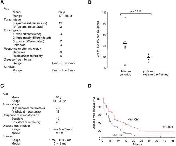
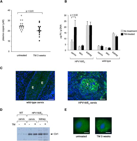
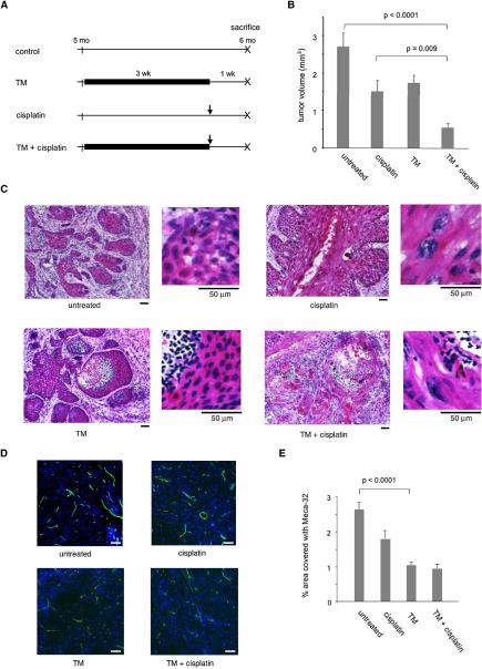
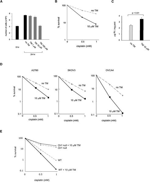
Uptake of the anticancer drug cisplatin is mediated by the copper transporter CTR1 in cultured cells. Here we show in human ovarian tumors that low levels of Ctr1 mRNA are associated with poor clinical response to platinum-based therapy. Using a mouse model of human cervical cancer, we demonstrate that combined treatment with a copper chelator and cisplatin increases cisplatin-DNA adduct levels in cancerous but not in normal tissues, impairs angiogenesis, and improves therapeutic efficacy. The copper chelator also enhances the killing of cultured human cervical and ovarian cancer cells with cisplatin. Our results identify the copper transporter as a therapeutic target, which can be manipulated with copper chelating drugs to selectively enhance the benefits of platinum-containing chemotherapeutic agents.


Figures

Figure 1
Figure 1. Low levels of Ctr1 mRNA in ovarian tumors are associated with poor clinical outcome
(A) Baseline characteristics of the 15 patients with advanced-stage epithelial ovarian carcinoma, whose tumors were analyzed for relative expression of the Ctr1 copper transporter. (B) Tumor Ctr1 mRNA levels in platinum sensitive and refractory/resistant ovarian cancer patients. Bars represent mean values. Ctr1 mRNA levels are expressed as a percentage relative to the internal control of human GUS mRNA. (C) Characteristics of 91 ovarian cancer patients from The Cancer Genome Atlas study. (D) Kaplan-Meier estimate of disease-free survival for 91 advanced-stage ovarian cancer patients according to Ctr1 expression levels. Patients whose tumors expressed Ctr1 mRNA at levels higher than the median value were classified as “High Ctr1” (red; 45 patients); the remaining patients were labeled “Low Ctr1” (blue; 46 patients). The vertical hash marks (at 7 and 10 months, High Ctr1) represent censored patients who were disease-free at the last follow-up time indicated. See also Figure S1.
Figure 2
Figure 2. Levels of Ctr1 mRNA and cisplatin adducts in various organs of the HPV16/E2 female mice
Organs were harvested from three six-month-old HPV16/E2 females and Ctr1 mRNA levels were determined by quantitative RT-PCR (hatched bars). Mouse L19 mRNA levels were used as internal controls. For measurement of cisplatin adducts, genomic DNA was purified from each organ (n=8) two hours after 6 mg/kg cisplatin was injected. Platinum was measured by inductively-coupled plasma mass spectrometry (ICP-MS) and normalized to the amount of DNA (solid bars). Error bars represent the standard error of the mean. See also Figure S2.
Figure 3
Figure 3. The copper chelator tetrathiomolybdate (TM) enhances cisplatin uptake into the cervix of HPV16/E2 mice
(A) Plasma copper levels in six-month-old HPV16/E2 females after a three-week daily treatment with 1 mg TM. Bars represent mean values. (B) Cisplatin adduct levels in the cervix, skin, and kidney of six-month-old HPV16/E2 females (n=12) and wild-type females (n=5) that received either the three-week TM treatment (solid bars) or no treatment (hatched bars). 6 mg/kg cisplatin was injected two hours before organs were harvested. Adduct levels were determined as in Figure 2. Error bars represent the standard error of the mean. (C) Ctr1 immunohistochemistry reveals positive cells (green) in tumors (T) of the six-month-old HPV16/E2 cervix (right panel) but not in the epithelium (E) of the wild-type cervix (left panel). DAPI staining is shown in blue. The panels shown are representative images of twelve fields in three tissue sections per mouse collected from three mice. Scale bar: 100 μm. (D) Tissue Ctr1 protein levels are elevated in the neoplastic cervix, but unchanged in the context of TM treatment. Wild-type and HPV16/E2 mice were either treated with 1 mg TM daily for 3 weeks prior to being sacrificed at 6 months of age, or did not receive such treatment. 5 μg of membrane extracts from tissues were pooled from three mice and applied on each well. (E) Ctr1 protein (green) is predominantly localized in intracellular compartments in cervical carcinoma cells, and is unchanged by TM treatment. HPV16/E2 mice were either treated with 1 mg TM daily for 3 weeks prior to being sacrificed at 6 months of age, or did not receive such treatment. Tissue sections were immunostained with anti-Ctr1(green). Nuclei are stained with DAPI (blue). The images were collected using a 20x objective lens on a fluorescent microscope, and are representative of > 500 cells in 12 sections from 6 tumors. Scale bar: 5 μm.
Figure 4
Figure 4. Preclinical trials assessing therapeutic effects of the copper chelator tetrathiomolybdate (TM) in combination with cisplatin on de novo cervical carcinomas
(A) Schematic diagrams of each therapeutic arm. Cohorts of 14 female HPV16/E2 mice were subjected either to a three-week oral administration of 1 mg TM daily starting at five months of age, or to a single intraperitoneal injection of 6 mg/kg cisplatin at five and ¾ months of age, or to both. All the mice were sacrificed at six months of age. (B) Tumor volumes in the cervix of six-month-old HPV16/E2 mice from each treatment arm. Approximately 30 sections per mouse from cohorts of 14 female HPV16/E2 mice were analyzed as described in Methods. Error bars represent the standard error of the mean. (C) Representative H&E-stained cervices from each treatment arm, with the treatment indicated below each panel. Cells are shown at a higher magnification on the right. The magnification is scaled by a bar below each panel indicating 50 μm. In the non-treated cancerous cervix (top left), invasive squamous cell carcinomas contain large keratinized cells with hyperchromatic nuclei. In the cisplatin-treated cervix (top right), the cancer cells have more abundant cytoplasm that appears keratinized, with enlarged, bizarre-shaped nuclei. In the TM-treated cervix (bottom left), the center of a tumor mass is necrotic and filled with inflammatory cell infiltrates. The cervix treated with both TM and cisplatin (bottom right) shows histological features of the effects seen with both cisplatin and TM monotherapies: enlarged nuclei, necrotic centers, and inflammatory cell infiltrates. The panels shown are representative of 4 fields in 30 tissue sections collected from cohorts of 14 HPV16/E2 mice. (D), (E) Vascular density of tumors after treatment. 15 tumor sections from five mice per treatment arm were stained with a pan-endothelial cell antibody Meca-32, followed by FITC-conjugated secondary antibody to visualize tumor endothelial cells. Images were collected from 5-7 fields per section, and vascular density was determined using the Metamorph Angiogenesis Tube Formation program. Data are expressed as the percentage of area covered with Meca-32-positive tube structures (E). Error bars represent the standard error of the mean. Representative images of Meca-32 (green) staining in the cervix of control and TM-treated HPV16/E2 mice are shown in D. Nuclei are stained with DAPI (blue). Scale bar: 50 μm.
Figure 5
Figure 5. Effects of the copper chelator tetrathiomolybdate (TM) on cisplatin sensitivity in cultured mammalian cells
(A) Effect of TM on cell proliferation in human cervical carcinoma cells. SiHa cells were plated in triplicates and treated with 0, 1, 10, or 100 μM TM for 24 hours. Numbers of cells before and after the treatment are shown. Error bars represent the standard error of the mean. (B), (C) Effect of TM on cisplatin sensitivity and accumulation in human cervical carcinoma cells. SiHa cells were plated in triplicates and either pretreated with 10 μM TM or mock treated for 24 hours before a two-hour incubation with cisplatin. For cell survival (B), data are expressed as percentage of viable cells compared with control cultures not exposed to cisplatin. For determining cisplatin accumulation (C), cells were lysed and, after a centrifugation, the supernatant was used to determine the platinum content and protein concentration. Cellular platinum readings were normalized to protein concentration. Mean values of the triplicates are shown. Error bars represent the standard error of the mean. (D) TM increases cisplatin sensitivity in human ovarian cancer cells. Cells were plated in triplicates and either pretreated with 10 μM TM or mock treated for 6 hours (A2780), 12 hours (SKOV3) or 24 hours (OVCA4) before cisplatin treatment. Data are expressed as percentage of viable cells compared with control cultures not exposed to cisplatin. (E) TM increases cisplatin sensitivity in a Ctr1-dependent manner. Isogenic mouse embryonic fibroblasts that are either wild-type or homozygous for a knockout allele of Ctr1 were either pretreated with 10 μM TM or mock treated for 24 hours before a two-hour incubation with cisplatin. The percentage of viable cells compared with control cultures not exposed to cisplatin is shown.

Similar articles

Cited by

References

    1. Arbeit JM, Howley PM, Hanahan D. Chronic estrogen-induced cervical and vaginal squamous carcinogenesis in human papillomavirus type 16 transgenic mice. Proc. Natl. Acad. Sci, USA. 1996;93:2930–2935. - PMC - PubMed
    1. Atallah E, Flaherty L. Treatment of metastatic malignant melanoma. Curr. Treat. Options Oncol. 2005;6:185–93. - PubMed
    1. Brewer GJ, Merajver SD. Cancer therapy with tetrathiomolybdate: antiangiogenesis by lowering body copper – a review. Integr. Cancer Ther. 2002;1:327–337. - PubMed
    1. Brewer GJ. The use of copper-lowering therapy with tetrathiomolybdate in medicine. Expert Opin. Investig. Drugs. 2009;18:89–97. - PubMed
    1. Bull PC, Thomas GR, Rommens JM, Forbes JR, Cox DW. The Wilson disease gene is a putative copper transporting P-type ATPase similar to the Menkes gene. Nat. Genet. 1993;5:327–337. - PubMed

Publication types

MeSH terms