Comparative Study
. 1991 Jun;6(6):993-1007.
doi: 10.1016/0896-6273(91)90239-v.

Differential expression of the p65 gene family

Affiliations
Comparative Study

Differential expression of the p65 gene family

B Wendland et al. Neuron. 1991 Jun.

Abstract

The genome of the marine ray Discopyge ommata contains at least three p65-related genes. o-p65-A is 84% identical, o-p65-B is 78% identical, and o-p65-C is only 41% identical to a previously characterized rat p65. The cytoplasmic domain, particularly the two regions that are similar to the regulatory domain of protein kinase C, are most highly conserved. The three genes are expressed in different but overlapping patterns in the central nervous system. o-p65-A immunoreactivity is found predominantly in forebrain, cerebellum, and neuroendocrine cells, while o-p65-B immunoreactivity is predominantly localized to the spinal cord, brainstem, and midbrain. Many synaptic vesicle proteins are members of small gene families that are differentially expressed, resulting in several unique combinations of these molecules in specific brain regions.


Figures
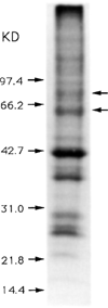
Figure 1
Figure 1. Discopyge Ommata Synaptic Vesicle Proteins
Cholinergic synaptic vesicles from the electric organ of D. ommata were purified and fractionated by SDS–PAGE. The gel was stained with Coomassie blue, revealing two vesicle-specific bands at 62 kd and 74 kd (arrows). These bands were sequenced directly or cleaved with CNBr, and some of the resulting fragments were sequenced.
Figure 2
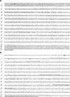
Figure 2. Nucleotide Sequences of Three p65 Related cDNAs
The nucleotide and predicted amino acid sequences for representatives of three p65 cDNAs are shown. (A), o-p65-A; (B), o-p65-B; and (C), o-p65-C. The region corresponding to the amino acid sequence determined from the CNBr cleavage fragments and amino terminus is underlined in o-p65-B. In-frame upstream stop codons are also underlined.
Figure 3
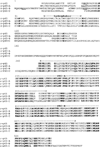
Figure 3. Alignment of Rat and Ray p65 Amino Acid Sequences
The amino acid sequences of the rat and three ray p65 proteins are aligned to maximize the extent of homology. Amino acid positions that are conserved in all four sequences are bold. Potential N-linked glycosylation sites are underlined. The hydrophobic domain and the two PKC-homologous regions are indicated above the sequence.
Figure 4
Figure 4. Schematic Representation of p65 Proteins
The four p65 sequences are depicted, and the homology to the rat sequence is indicated for the various domains. The two PKC-homologous regions are indicated (dark stippling), as is the hydrophobic domain (zig zags). Glycosylation sites are depicted (solid circles). The K in rat p65, o-p65-A, and o-p65-B indicates a lysine-rich region, and the H in o-p65-C indicates a histidine-rich region. An insert and a region of low homology in o-p65-C relative to the other sequences is shaded (light stippling). An amino-terminal extension of o-p65-B and a carboxy-terminal extension of o-p65-C are depicted.
Figure 5
Figure 5. Differential Expression of the p65 Gene Family
Northern blots of RNA from D. ommata tissues are shown. (A) Poly(A)+ RNA probed with o-p65-A. Lane 1, spinal cord; lane 2, electric lobe; lane 3, forebrain. (B) Poly(A)+ RNA probed with o-p65-B. Lane 1, heart; lane 2, liver; lane 3, muscle; lane 4, total brain; lane 5, spinal cord; lane 6, electric organ; lane 7, electric lobe; and lane 8, forebrain. (C) Total RNA probed with o-p65-C. Lane 1, electric lobe; lane 2, liver; and lane 3, forebrain. Blots were stained with methylene blue (A-C) and probed for actin message (B) to confirm presence of intact RNA.
Figure 6
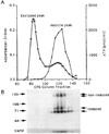
Figure 6. Immunoblot Analysis of the p65 Immunoreactive Species Copurifying with Electric Organ Vesicles
(A) Graph illustrating the final stage of vesicle isolation: CPG chromatography. Open squares indicate absorbance at 310 nm, and solid diamonds indicate ATP. (B) Immunoblot in which an equal volume of every fifth fraction from 75–145 of the same prep has been fractionated by SDS-PAGE, blotted to nitrocellulose, and probed with the anti-62 kd antibodies or antibodies to the synaptic vesicle protein, VAMP (Trimble et al., 1988). Molecular weight markers in kilodaltons and the location of VAMP are indicated on the left. The positions of the reduced and unreduced forms of the immunolabeled bands are indicated on the right.
Figure 7
Figure 7. Immunoblot Analysis of o-p65-A and o-65-B in the Ray CNS
Total protein (40 µg) from various regions of the CNS was fractionated on a 10% SDS-PAGE gel and probed with MAb 48 to detect o-p65-A (A) or with anti-62 kd antibodies to detect o-p65-B (B). Lane 1, vesicles (4.5 µg); lane 2, organ; lane 3, spinal cord; lane 4, brainstem; lane 5, cerebellum; lane 6, midbrain; lane 7, forebrain. Lanes 8 and 9 in (B) are muscle and liver, respectively.
Figure 8
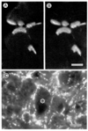
Figure 8. o-p65-B Immunoreactivity in Nerve Terminals
(A) Indirect immunofluorescence localization of o-p65-B immunoreactivity in a 15 µm cross section of ray muscle fibers. The secondary antibody is fluorescein isothiocyanate–labeled. (B) Same field stained with rhodamine-labeled α-bungarotoxin to reveal neuromuscular junctions.(C) o-p65-B immunoreactivity in the ray brainstem. Immunofluorescence staining of 6 µm frozen section. Note the brightly stained nerve terminals outlining the soma of the large neuron in the center (asterisk). Bars: 40 µm (A and B); 100 µm (C).
Figure 9
Figure 9. Immunofluorescence Staining of o-p65-A and o-65-B in the Ray CNS: Comparison with SV2
Shown are 7 µm frozen sections of spinal cord (A–C), cerebellum (D–F), midbrain (G–I), and forebrain (J–L). (A, D, G, and J) SV2. (B, E, H, and K) o-p65-A. (C, F, I, and L) o-p65-B. The molecular (M) and granule cell (G) layers of the cerebellum are indicated. Bars: 40 µm (A–D; F–I); 20 µm (E); 100 µm (J–L). The section in (L) was exposed longer than comparable sections, but the low amount of punctate staining demonstrates the paucity of o-p65-B immunoreactive terminals in the forebrain.
Figure 10
Figure 10. Immunofluorescence Staining of o-p65-A and o-65-B in Neuroendocrine Cells
Shown are 7 µm frozen sections of anterior pituitary (A–C) and pancreas (B–F). (A and D) SV2. (B and E) o-p65-A. (C and F) o-p65-B. The staining around the dark spaces in (A) and (B) represents neuroendocrine cells lining the capillary passages. Bar, 40 µm. The distribution and arrangement of the SV2- and p65-positive cells in the ray pituitary and pancreas are consistent with previous studies of the elasmobranch endocrine system (Thomas, 1940; Hoar and Randall, 1969).
Figure 11
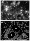
Figure 11. Immunofluorescence Staining of p65-A in Rat Shown are 7 µm frozen sections of rat spinal cord (ventral horn)
(A) p65-A and (B) SV2. Two neurons surrounded by SV2 immuno-reactive terminals are indicated with asterisks. Bar, 100 µm. The several large fluorescent spots in (A) are background and not consistently observed.

Similar articles

Cited by

References

    1. Almers W. Exocytosis. Annu. Rev. Physiol. 1990;52:607–624. - PubMed
    1. Baumert M, Maycox PR, Navone F, De Camilli P, Jahn R. Synaptobrevin: an integral membrane protein of 18000 daltons present in small synaptic vesicles of rat brain. EMBO J. 1989;8:379–384. - PMC - PubMed
    1. Baumert M, Takei K, Hartinger J, Burger PM, Fischer von Mollard G, Maycox PR, De Camilli P, Jahn R. P29: a novel tyrosine-phosphorylated membrane protein present in small clear vesicles of neurons and endocrine cells. J. Cell Biol. 1990;110:1285–1294. - PMC - PubMed
    1. Bourne HR. Do GTPases direct membrane traffic in secretion? Cell. 1988;53:669–671. - PubMed
    1. Buckley K, Kelly RB. Identification of a transmembrane glycoprotein specific for secretory vesicles of neural and endocrine cells. J. Cell Biol. 1985;700:1284–1294. - PMC - PubMed

Publication types

Associated data

LinkOut - more resources