. 2010 Jun 11;38(5):637-48.
doi: 10.1016/j.molcel.2010.04.017.

A ubiquitin-binding domain in Cockayne syndrome B required for transcription-coupled nucleotide excision repair

Affiliations

A ubiquitin-binding domain in Cockayne syndrome B required for transcription-coupled nucleotide excision repair

Roy Anindya et al. Mol Cell. .

Abstract

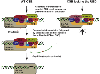
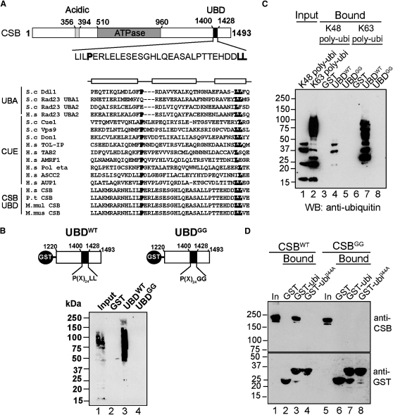
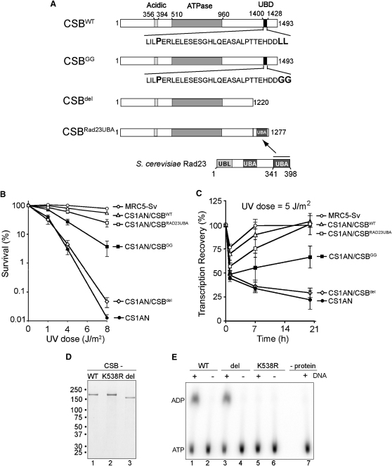
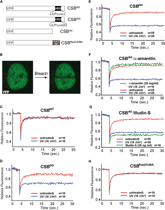
Transcription-coupled nucleotide excision repair (TC-NER) allows RNA polymerase II (RNAPII)-blocking lesions to be rapidly removed from the transcribed strand of active genes. Defective TCR in humans is associated with Cockayne syndrome (CS), typically caused by defects in either CSA or CSB. Here, we show that CSB contains a ubiquitin-binding domain (UBD). Cells expressing UBD-less CSB (CSB(del)) have phenotypes similar to those of cells lacking CSB, but these can be suppressed by appending a heterologous UBD, so ubiquitin binding is essential for CSB function. Surprisingly, CSB(del) remains capable of assembling nucleotide excision repair factors and repair synthesis proteins around damage-stalled RNAPII, but such repair complexes fail to excise the lesion. Together, our results indicate an essential role for protein ubiquitylation and CSB's UBD in triggering damage incision during TC-NER and allow us to integrate the function of CSA and CSB in a model for the process.


Figures

None
Graphical abstract
Figure 1
Figure 1
Identification of a Ubiquitin-Binding Domain in CSB (A) Upper: Schematic drawing of CSB indicating the UBD. Lower: Multiple sequence alignment of UBA and CUE domains from various proteins. Conserved residues that contribute to the hydrophobic core are in bold. Boxes at the top denote the locations of α helices in the yeast Cue2 CUE1 and human HHR23A UBA1 domains, respectively. (B) Upper: GST-UBDWT and GST-UBDGG fusion proteins used for ubiquitin binding assay. Lower: Ubiquitylated proteins from human cell lysates retained on immobilized GST, GST-UBDWT, or GST-UBDLL. Total lysate (5%) and eluted proteins were analyzed by SDS-PAGE, followed by anti-ubiquitin immunoblotting. (C) Pure multiubiquitin chains retained on immobilized GST, GST-UBDWT, or GST-UBDGG, analyzed as in (B). (D) Binding of CSBWT or CSBGG to immobilized GST, GST-ubiquitin, or GST-ubiquitinI44A. Total human cell lysate (10%) and eluted proteins were analyzed by SDS-PAGE, followed by anti-CSB immunoblotting (upper panel). Equivalent loading was examined by anti-GST immunoblotting (lower panel). See also Figure S1.
Figure 2
Figure 2
Functional Importance of CSB's Ubiquitin-Binding Domain (A) Schematic representation of proteins stably expressed in CSB-deficient (CS1AN-sv) human fibroblasts. (B) UV-survival experiment, with percentage surviving cells (logarithmic scale) plotted against UV dose. Error bars indicate standard error based on three independent experiments. (C) RNA synthesis after UV irradiation measured as the relative incorporation of 3H-uridine in 5 J/m2-irradiated cells compared with unirradiated cells (100%). Relative transcription is plotted against the UV dose. Error bars indicate standard error based on three independent experiments. (D) SDS-PAGE analysis of overexpressed CSB proteins, purified from human cells, stained with Coomassie blue. Migration of molecular weight markers is indicated on the left. (E) DNA-dependent ATPase activity of CSB proteins, measured as generation of α-P32-ADP from α-P32-ATP. CSB K538R is used as negative control. This mutation, in the invariant lysine residue in the NTP-binding motif of CSB, inhibits ATP hydrolysis (Citterio et al., 1998). See also Figure S2.
Figure 3
Figure 3
Association of CSB and RNAPII with the DHFR Promoter (A) Upper: Schematic of DHFR and position of PCR primers. Lower: Kinetics of CSB occupancy at the DHFR promoter after UV irradiation measured by ChIP. The results obtained in untreated cells were set to one, and the other values relative to that, with standard deviation. Data are representative of three independent experiments. (B) As in (A) but for RNAPII.
Figure 4
Figure 4
CSB Lacking the UBD Becomes Immobilized at DNA Damage in a TC-NER-Dependent Manner (A) Scheme of the different constructs used. (B) Confocal images of a human fibroblast stably expressing YFP-CSBdel before and after bleaching of a strip along the nucleus width. (C-E) Strip-FRAP graphs showing the mobility of (C) YFP-CSBWT, (D) YFP-CSBGG, and (E) YFP-CSBdel in untreated (red) and UV-irradiated cells (blue). (F) As in (E), but using cells pre-treated with α-amanitin to block transcription (green). (G) As in (E), but also using cells treated with 30 ng/ml Illudin S (green). (H) Strip-FRAP graph showing the mobility of YFP-CSBRad23UBA in untreated (red) and UV-irradiated cells (blue). See also Figure S3.
Figure 5
Figure 5
CSB Lacking the UBD Remains Capable of Assembling TC-NER Complexes after DNA Damage (A) Western blot of CSB-specific ChIPs using antibodies directed against the factors indicated on the left, in cell lines expressing CSB or CSBdel, as indicated. (B and C) As in (A), but with antibodies against proteins involved in repair synthesis (B), or ubiquitylated histone H2A (C). Histone proteins in lower panels serve as loading controls.
Figure 6
Figure 6
CSB Lacking the UBD Fails to Support TC-NER Damage Incision (A) Upper: Schematic of the TC-NER assay (Spivak et al., 2006). Lower: Stylized example of result from normal TC-NER reaction. As long as damages persist in a fragment, T4 EndoV will digest it (+), resulting in a smaller amount of fragment compared with the untreated control lanes (−). (B) Expected result from assay if damage incision (one or both) is defective in the tested cell line. (C) TC-NER in endogenous DHFR gene in the cell lines indicated immediately below blots probed with a strand-specific probe. TS, transcribed strand; NTS, nontranscribed strand.

Comment in

Similar articles

Cited by

References

    1. Anindya R., Aygun O., Svejstrup J.Q. Damage-induced ubiquitylation of human RNA polymerase II by the ubiquitin ligase Nedd4, but not Cockayne syndrome proteins or BRCA1. Mol. Cell. 2007;28:386–397. - PubMed
    1. Bergink S., Salomons F.A., Hoogstraten D., Groothuis T.A., de Waard H., Wu J., Yuan L., Citterio E., Houtsmuller A.B., Neefjes J. DNA damage triggers nucleotide excision repair-dependent monoubiquitylation of histone H2A. Genes Dev. 2006;20:1343–1352. - PMC - PubMed
    1. Bhatia P.K., Verhage R.A., Brouwer J., Friedberg E.C. Molecular cloning and characterization of Saccharomyces cerevisiae RAD28, the yeast homolog of the human Cockayne syndrome A (CSA) gene. J. Bacteriol. 1996;178:5977–5988. - PMC - PubMed
    1. Bienko M., Green C.M., Crosetto N., Rudolf F., Zapart G., Coull B., Kannouche P., Wider G., Peter M., Lehmann A.R. Ubiquitin-binding domains in Y-family polymerases regulate translesion synthesis. Science. 2005;310:1821–1824. - PubMed
    1. Chen L., Shinde U., Ortolan T.G., Madura K. Ubiquitin-associated (UBA) domains in Rad23 bind ubiquitin and promote inhibition of multi-ubiquitin chain assembly. EMBO Rep. 2001;2:933–938. - PMC - PubMed

Publication types

MeSH terms