. 2010 Jun 11;38(5):712-21.
doi: 10.1016/j.molcel.2010.05.024.

The AAA-ATPase Rea1 drives removal of biogenesis factors during multiple stages of 60S ribosome assembly

Affiliations

The AAA-ATPase Rea1 drives removal of biogenesis factors during multiple stages of 60S ribosome assembly

Jochen Bassler et al. Mol Cell. .

Abstract

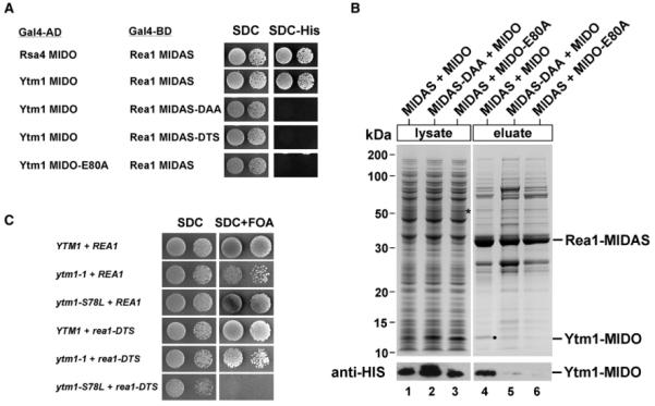
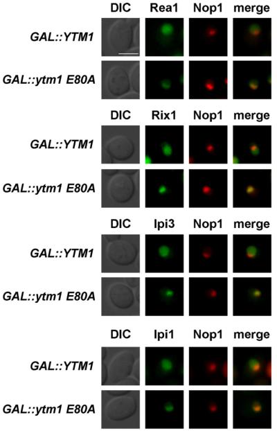
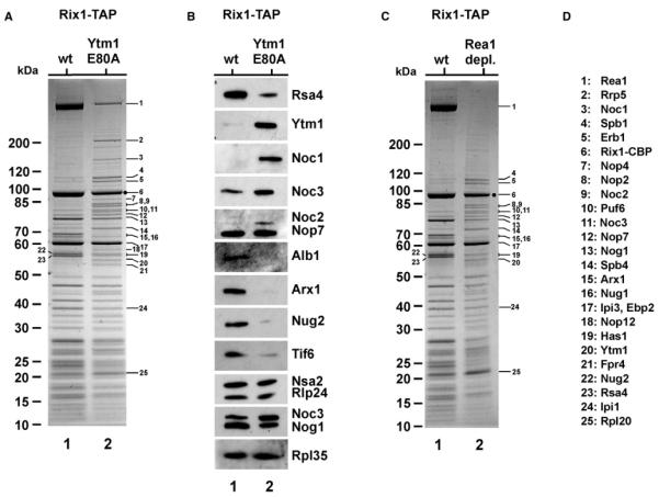
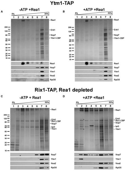
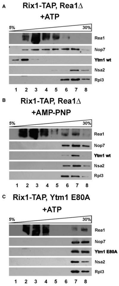
The AAA(+)-ATPase Rea1 removes the ribosome biogenesis factor Rsa4 from pre-60S ribosomal subunits in the nucleoplasm to drive nuclear export of the subunit. To do this, Rea1 utilizes a MIDAS domain to bind a conserved motif in Rsa4. Here, we show that the Rea1 MIDAS domain binds another pre-60S factor, Ytm1, via a related motif. In vivo Rea1 contacts Ytm1 before it contacts Rsa4, and its interaction with Ytm1 coincides with the exit of early pre-60S particles from the nucleolus to the nucleoplasm. In vitro, Rea1's ATPase activity triggers removal of the conserved nucleolar Ytm1-Erb1-Nop7 subcomplex from isolated early pre-60S particle. We suggest that the Rea1 AAA(+)-ATPase functions at successive maturation steps to remove ribosomal factors at critical transition points, first driving the exit of early pre-60S particles from the nucleolus and then driving late pre-60S particles from the nucleus.


Figures

Figure 1
Figure 1. The MIDO of Ytm1 Interacts with Rea1’s MIDAS In Vivo and In Vitro
(A) Yeast two-hybrid interaction between wild-type and mutant alleles of Rea1-MIDAS and Ytm1-MIDO. Yeast two-hybrid plasmids expressing the indicated GAL4-BD (GAL4 DNA-binding domain) and GAL4-AD (GAL4 activation domain) fusion proteins were transformed into the yeast reporter strain PJ69-4A. Transformants were spotted in 10-fold serial dilutions onto SDC-Trp-Leu (SDC) or SDC-Trp-Leu-His (SDC-His) and incubated at 30 °C for 4 days. The Rea1-MIDAS comprises residues 4622–4910; the MIDO of Rsa4, residues 1–154; and the MIDO of Ytm1, residues 1–92. Growth on SDC-His plate indicates a two-hybrid interaction. (B) Rea1-MIDAS and Ytm1-MIDO bind directly to each other. The GST-TEV-tagged MIDAS of Rea1 (wild-type or DAA mutant; residues 4608–4910) was coexpressed with His6-Ytm1-MIDO (1–92 aa) or the His6-Ytm1-MIDO E80A mutant in E. coli in the indicated combinations. The GST-MIDAS fusion proteins were affinity purified on GSH beads and eluted by TEV cleavage. Supernatants and eluates were analyzed by SDS-PAGE and Coomassie staining (top) and western blotting (bottom) using anti-HIS antibodies to detect Ytm1 in eluates and total cell lysates. Protein bands above GST-MIDAS are E. coli contaminants. (C) The ytm1-MIDO mutant S78L is genetically linked to the rea1-DTS mutant. The YTM1Δ/REA1Δ double-shuffle strain was transformed with wild-type and the indicated mutant alleles of YTM1 and REA1, respectively. Transformants were spotted in 10-fold serial dilutions onto SDC-Trp-Leu (SDC) or SDC+FOA to test for synthetic lethality. Plates were incubated at 30 °C for 2 (SDC) or 4 days (SDC+FOA). A sequence analysis of Ytm1 MIDO is shown in Figure S1.
Figure 2
Figure 2. Dominant-Lethal Ytm1 E80A Inhibits Formation of 60S Subunits
(A) The ytm1 E80A mutant causes a dominant-negative growth phenotype. Wild-type YTM1 and the indicated ytm1 E80A mutant allele were overexpressed under the control of the inducible GAL1 promoter in a wild-type yeast strain. Transformants were spotted in 10-fold serial dilutions on SDC-Leu (glucose) and SGC-Leu (galactose) plates. Glucose plates were incubated for 2 days and galactose plates for 7 days at 30 °C. (B) Overexpression of the Ytm1 E80A protein blocks 27S to 7S pre-rRNA processing. Cells were grown in SRC-Leu (time point 0) before expression of YTM1, ytm1 E80A, RSA4, rsa4 E114D was induced by transferring cells into SGC-Leu. Total RNA was isolated after 0, 2, 4, 6, and 8 hr of induction and analyzed by northern blot. Probes used for the shown autoradiographs are indicated aside. Panels for 35S, 27SA2, and 23S rRNA show detection from the same exposure time. (C) Analysis of 60S ribosomal export in the dominant-negative GAL::ytm1 E80A mutant. Wild-type yeast strain, harboring plasmids pRS314-RFP-NOP1-RPL25-GFP and YCplac111-GAL::YTM1 wild-type or ytm1 E80A, was grown (30 °C) in raffinose-containing medium before cells were shifted to galactose medium to induce overexpression of Ytm1 and Ytm1 E80A, respectively. The subcellular location of the Rpl25-GFP (reporter for 60S export) and mRFP-Nop1 (nucleolar marker) were analyzed by fluorescence microscopy after 5 hr shift. Single and merged pictures of Rpl25-GFP and mRFP-Nop1 as well as Nomarski (DIC) pictures, are shown. Scale bar, 2 μm. For further characterization of the ytm1 E80A mutant, see Figure S2.
Figure 3
Figure 3. GFP-Rea1 and the Rix1-Ipi3-Ipi1 Subcomplex Become Mislocalized upon Overexpression of Dominant-Negative Ytm1 E80A
Yeast strains carrying chromosomal GFP fusion proteins (PRea1::GFP-Rea1, Rix1-GFP, Ipi3-GFP, and Ipi1-GFP) were transformed with pRS314 mRFP-Nop1 (nucleolar marker) and YCplac111-GA-L::YTM1 or YCplac111-GAL::ytm1-E80A. Transformants were grown in SRC-Leu-Trp medium (raffinose) and shifted to SGC-Leu-Trp (galactose) medium for 6 hr to induce overexpression of Ytm1 or Ytm1-E80A. Subcellular location of GFP fusion proteins was determined by fluorescence microscopy. Nomarski (DIC), GFP, RFP, and merge pictures are shown. Scale bar, 2μm. Figure S3 shows the localization of additional 60S biogenesis factors upon Ytm1 E80A overexpression.
Figure 4
Figure 4. Rix1-TAP Co-Enriches a Nucleolar Pre-60S Particle upon Overexpression of Ytm1 E80A or Rea1 Depletion
(A and B) Yeast strain Rix1-TAP was transformed with YCplac111-GAL::YTM1 or YCplac111-GAL::ytm1 E80A. Transformants were grown in SRC-Leu medium (raffinose) and shifted to YPG (galactose) medium for 6 hr to induce overexpression of Ytm1 or Ytm1 E80A. Subsequently, TAP purifications were performed, and final EGTA eluates were analyzed by 4%–12% gradient SDS-PAGE and Coomassie staining (A) and western blotting (B) using the indicated antibodies. Molecular weight marker (M) is indicated. Labeled protein bands were identified by mass spectrometry (see D for band assignation). (C) Yeast strain Rix1-TAP with GAL::HA-REA1 was grown in YPG (galactose) and shifted to YPD (glucose) for 16 hr to deplete for Rea1. Subsequently, Rix1-TAP purification was performed, and the final EGTA eluate were analyzed by 4%–12% gradient SDS-PAGE and Coomassie staining (Rea1 depl., lane 2). This eluate was compared to a Rix1-TAP purification when Rea1 was overexpressed (grown in YPG, “wt” lane 1). Molecular weight marker (M) is indicated, and labeled protein bands were identified by mass spectrometry (see D for band assignation). (D) Band assignation. The rRNA content of the purified particles, as well as the rRNA processing defect of a Gal::REA1 strain, is shown in Figure S4.
Figure 5
Figure 5. Rea1 Extracts Ytm1-Erb1-Nop7 from the Purified Pre-60S Ribosomal Particle In Vitro
(A and B) The Ytm1-TAP particle was affinity purified, mixed with purified Rea1, and incubated for 45 min at 23 °C with ± 4 mM ATP. Subsequently, the mixture was loaded on a 5%–30% sucrose gradient and centrifuged for 16 hr at 27.000 rpm. Gradient fractions 1–8 were analyzed by SDS-PAGE and Coomassie staining (top) or western blotting using the indicated antibodies (bottom). (C and D) The Rix1-TAP particle was affinity purified from Rea1-depleted cells (see Figure 4C), mixed with purified Rea1, and incubated for 45 min at 23 °C with ± 4 mM ATP. Subsequently, the mixture was loaded on a 5%-30% sucrose gradient and centrifuged for 16 hr at 27.000 rpm as described in (A) and (B). Gradient fractions 1–8 were analyzed by SDS-PAGE and silver staining (top) or western blotting using the indicated antibodies (bottom). Bands released by ATP treatment were Erb1 (•) and Ytm1 (*), which were identified by mass spectroscopy.
Figure 6
Figure 6. The Release of Ytm1 Depends on the Ytm1-Rea1 Interaction and ATP Hydrolysis
(A–C) The Rix1-TAP particle was affinity purified from Rea1-depleted cells (A and B) (see Figure 4C) or from Ytm1-E80A-induced cells (C) (see Figure 4A), mixed with purified Rea1, and incubated for 45 min at 23 °C with 4 mM ATP or AMP-PNP, respectively. Subsequently, the mixture was loaded on a 5%–30% sucrose gradient and centrifuged for 16 hr at 27.000 rpm. Gradient fractions 1–8 were analyzed by western blotting using the indicated antibodies.

Similar articles

Cited by

References

    1. Bassler J, Grandi P, Gadal O, Lessmann T, Petfalski E, Tollervey D, Lechner J, Hurt E. Identification of a 60S preribosomal particle that is closely linked to nuclear export. Mol. Cell. 2001;8:517–529. - PubMed
    1. Bassler J, Kallas M, Hurt E. The NUG1 GTPase reveals and N-terminal RNA-binding domain that is essential for association with 60 S pre-ribosomal particles. J. Biol. Chem. 2006;281:24737–24744. - PubMed
    1. Davies JM, Brunger AT, Weis WI. Improved structures of full-length p97, an AAA ATPase: implications for mechanisms of nucleotide-dependent conformational change. Structure. 2008;16:715–726. - PubMed
    1. de la Cruz J, Sanz-Martínez E, Remacha M. The essential WD-repeat protein Rsa4p is required for rRNA processing and intranuclear transport of 60S ribosomal subunits. Nucleic Acids Res. 2005;33:5728–5739. - PMC - PubMed
    1. DeLaBarre B, Brunger AT. Nucleotide dependent motion and mechanism of action of p97/VCP. J. Mol. Biol. 2005;347:437–452. - PubMed

Publication types

MeSH terms

Substances