. 2010 Jun 11;38(5):733-45.
doi: 10.1016/j.molcel.2010.05.001.

Monoubiquitination of RPN10 regulates substrate recruitment to the proteasome

Affiliations

Monoubiquitination of RPN10 regulates substrate recruitment to the proteasome

Marta Isasa et al. Mol Cell. .

Abstract

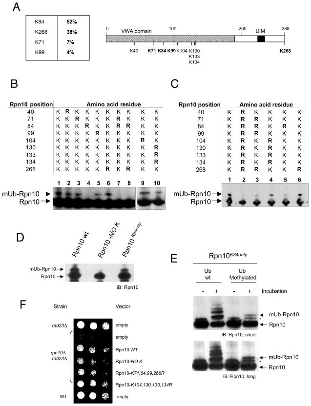
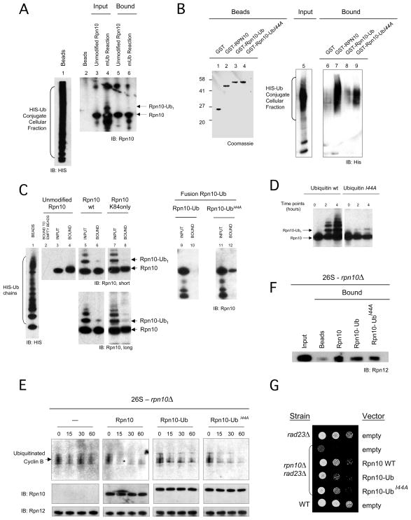
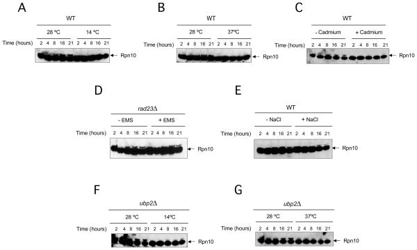
The proteasome recognizes its substrates via a diverse set of ubiquitin receptors, including subunits Rpn10/S5a and Rpn13. In addition, shuttling factors, such as Rad23, recruit substrates to the proteasome by delivering ubiquitinated proteins. Despite the increasing understanding of the factors involved in this process, the regulation of substrate delivery remains largely unexplored. Here we report that Rpn10 is monoubiquitinated in vivo and that this modification has profound effects on proteasome function. Monoubiquitination regulates the capacity of Rpn10 to interact with substrates by inhibiting Rpn10's ubiquitin-interacting motif (UIM). We show that Rsp5, a member of NEDD4 ubiquitin-protein ligase family, and Ubp2, a deubiquitinating enzyme, control the levels of Rpn10 monoubiquitination in vivo. Notably, monoubiquitination of Rpn10 is decreased under stress conditions, suggesting a mechanism of control of receptor availability mediated by the Rsp5-Ubp2 system. Our results reveal an unanticipated link between monoubiquitination signal and regulation of proteasome function.


Figures

Figure 1
Figure 1. Rpn10 is monoubiquitinated in vivo
(A) Logarithmically growing wild-type yeast cells (see strain list in Table S1) analyzed by western blotting using Rpn10 antibody. Time points were taken as shown. (B) Purified TAP-tagged Rpn10, analyzed by western blotting. Increasing amounts of fractions were visualized by short (left) and long film exposures. Bands ‘a’ and ‘b’ represent putative mono and diubiquitinated Rpn10. Asterisk, Rpn10 breakdown product. (C) Native 6xHis-Ubiquitin conjugates eluted at different imidazole concentrations (lanes 1 to 7), analyzed by western blotting with Rpn10 antibody. The same purification and analysis procedure was performed using a hul5Δ strain (lanes 8 to 14). Immunoblots with antibodies against proteasome subunits Rpt1, α7 and Rpn12 are included. Asterisks, unspecific bands detected with Rpn10 antibody.
Figure 2
Figure 2. Monoubiquitination of proteasomal Rpn10 is dynamically regulated
(A–B) Fraction UR8 (see supplemental data and Figure S2A) was incubated without ATP (lanes 1 to 4), with 5mM ATP (lanes 5 to 8) or with 5 mM ATP and purified proteasomes (lanes 10 to 13), at 30 °C, and time points were taken. In B, assays were performed using UR8 fraction and proteasomes from hul5Δ strains. (C) Total cell lysates from a wild-type strain were applied to a Superose 6 column. Fractions obtained were analyzed by western blotting of Rpn10, Rpn12 or α7 proteasome components. (D) Proteasomes purified in the presence of 5 mM ATP and 5 μM of MG132 were analyzed by two dimensioned electrophoresis (isoelectrofocusing and 4–12% gradient SDS-PAGE) followed by Rpn10 immunodetection. Asterisk, Rpn10 breakdown product.
Figure 3
Figure 3. Levels of monoubiquitinated Rpn10 are controlled by Rsp5-Ubp2 enzymatic system in vivo
(A) Wild-type, rsp5-1, ubp2Δ and double rsp5-1 ubp2Δ strains, carrying GST-Rpn10 galactose inducible plasmid (plasmids are listed in Table S2), were expressed and shifted to restrictive temperature. GST-Rpn10 was pulled down and analyzed by Rpn10 western blotting. Lanes 1 to 4, cultures grown at 28 °C. Lanes 5 to 8, cultures grown at 35 °C. Induction levels are shown in Figure S3B. (B) Monoubiquitinated Rpn10 from a ubp6Δ strain (see Figure S3E and supplemental data) was incubated with equimolar amounts of Ubp2, Ubp6 and Ubp6C118A. Reactions were analyzed by Rpn10 western blotting. (C) Total cell lysates from wild-type, ubp2Δ and ubp6Δ strains were applied to a Superose 6 chromatography. Fractions that contain eluted proteasome were analyzed by Rpn10 western blotting.
Figure 4
Figure 4. Reconstitution of the reaction of Rpn10 monoubiquitination in vitro
(A) Recombinant Rsp5, Rpn10, E1, Ubc4 and ubiquitin were incubated as indicated. Right lanes show a reaction in which Rpn10 input was doubled. Lower panel, longer exposures which show diubiquitinated Rpn10 (Rpn10-Ub2) more clearly. (B) Proteasomes (wild-type and Rpn10UIM) were incubated with Rsp5, E1, Ubc4 and ubiquitin. (C) Proteasomes (wild-type and Rpn10UIM) were incubated with a cell lysate fraction. An inhibitory effect of o-phenanthroline was observed when 1 mM final concentration of this compound was added to the reaction. Asterisk, Rpn10 breakdown product. (D–E) Scaled-up reactions of Rpn10 monoubiquitination in vitro, using wild-type (D) or methylated ubiquitin (E). Asterisk, unspecific band also observed in 4A. (F) Autoubiquitinated Rsp5 using wild-type or methylated ubiquitin, from reactions similar to (D) and (E), respectively.
Figure 5
Figure 5. Analysis of Rpn10 lysines modified by ubiquitin
(A) Summary of mass spectrometry analysis of Rpn10. Left, relative abundance of the Gly-Gly peptides found. Right, scheme of Rpn10 protein including all lysines contained in the sequence. Modified lysines are shown in bold. (B–D) GST-Rpn10 K to R mutants including all Rpn10 lysines (oligos are listed in Table S3) were expressed under galactose induction. Pulled down GST-Rpn10 forms were analyzed by Rpn10 western blotting. (E) Monoubiquitination reactions of Rpn10K84only mutant using wild-type and methylated ubiquitin. Asterisk, electrophoretic artifact shown by the mutant, observed also in non-incubated samples. Short and long exposures of the film are shown. (F) Ability of a set of Rpn10 mutants to rescue growth defect exhibited by rpn10Δrad23Δ yeast strain (see supplemental data). Rpn10 forms were expressed at 22 °C using a galactose inducible vector, selected with a URA3 marker. Cells (3×10⁴) were spotted in the first column and 3/7 serial dilutions were made for the successive columns.
Figure 6
Figure 6. Monoubiquitinated Rpn10 shows an inactive UIM and decreases the proteolytic activity of the proteasome
(A) Binding assay of Rpn10 and mUb-Rpn10 to a fraction of endogenous HIS-ubiquitin conjugates (see Figure S5A and supplemental data) immobilized on Ni-NTA beads (lane 1). Liquid phase inputs include unmodified Rpn10, as a control (lane 3), and a monoubiquitination reaction sample (mUb reaction), which contains both unmodified Rpn10 and mUb-Rpn10 (Rpn10-Ub1) (lane 4). Bound material is shown in lanes 5 and 6. (B) Binding assay of Rpn10 and C-terminal ubiquitin fusions (Rpn10-Ub and Rpn10-UbI44A), immobilized in beads (lanes 1 to 4), to a fraction of endogenous HIS-ubiquitin conjugates (input, lane 5). Bound material was eluted and analyzed by HIS western blotting (lanes 6 to 9). (C) Binding assay of unmodified and mUb-Rpn10 (wild-type and K84only mutant), Rpn10-Ub and Rpn10-UbI44A to unanchored polyubiquitin chains immobilized in beads (lane 1). Input and bound material are shown for each sample (lanes 3 to 12). A control of Rpn10 binding to empty beads is included in lane 2. A longer film exposure is shown for lanes 5 to 8. (D) Reaction of Rpn10 monoubiquitination in vitro using wild-type ubiquitin and I44A mutant. (E) Time-course degradation assays in vitro with proteasomes deficient in Rpn10, equimolar amounts of Rpn10 forms and ubiquitinated cyclin B. Points at indicated times were taken and analyzed by anti cyclin B, Rpn10 and Rpn12 western blotting. (F) Rpn10-Ub binding to rpn10Δ proteasomes. GST-Rpn10, GST-Rpn10-Ub and GST-Rpn10-UbI44A were immobilized to beads and proteasomes were applied as the soluble phase. (G) Rescue of Rpn10 function by Rpn10-Ub forms, including Rpn10, Rpn10-Ub and Rpn10-UbI44A expressed from a vector. The assay was performed as in figure 5F. Levels of protein expression are shown in Figure S5D.
Figure 7
Figure 7. Monoubiquitination of Rpn10 is decreased under certain stress conditions
(A–G) Yeast cells (wild-type, rad23Δ and ubp2Δ) were grown in YPD liquid media at the described conditions. Cadmium: 200 μM, Ethyl methyl sulfoxide (EMS): 0.08%, NaCl: 500 mM. Samples were taken at indicated times, number of cells normalized and analyzed by western blotting with Rpn10 antibody.

Comment in

References

    1. Chen L, Madura K. Rad23 promotes the targeting of proteolytic substrates to the proteasome. Mol Cell Biol. 2002;22:4902–4913. - PMC - PubMed
    1. Ciechanover A, Brundin P. The ubiquitin proteasome system in neurodegenerative diseases: sometimes the chicken, sometimes the egg. Neuron. 2003;40:427–446. - PubMed
    1. Crosas B, Hanna J, Kirkpatrick DS, Zhang DP, Tone Y, Hathaway NA, Buecker C, Leggett DS, Schmidt M, King RW, Gygi SP, Finley D. Ubiquitin chains are remodeled at the proteasome by opposing ubiquitin ligase and deubiquitinating activities. Cell. 2006;127:1401–1413. - PubMed
    1. Deveraux Q, Ustrell V, Pickart C, Rechsteiner M. A 26 S protease subunit that binds ubiquitin conjugates. J Biol Chem. 1994;269:7059–7061. - PubMed
    1. Di Fiore PP, Polo S, Hofmann K. When ubiquitin meets ubiquitin receptors: a signalling connection. Nat Rev Mol Cell Biol. 2003;4:491–497. - PubMed

Publication types

MeSH terms

Substances