

Impact of developmental lead exposure on splenic factors
- PMID: 20542052
- PMCID: PMC3219921
- DOI: 10.1016/j.taap.2010.06.003
Impact of developmental lead exposure on splenic factors
Abstract
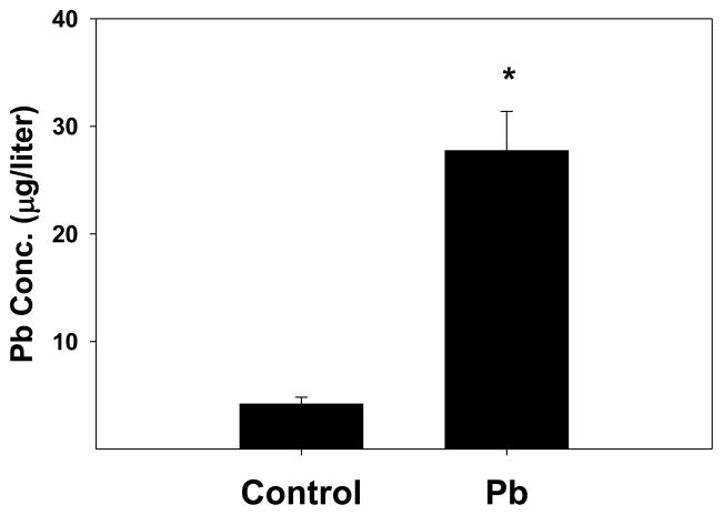
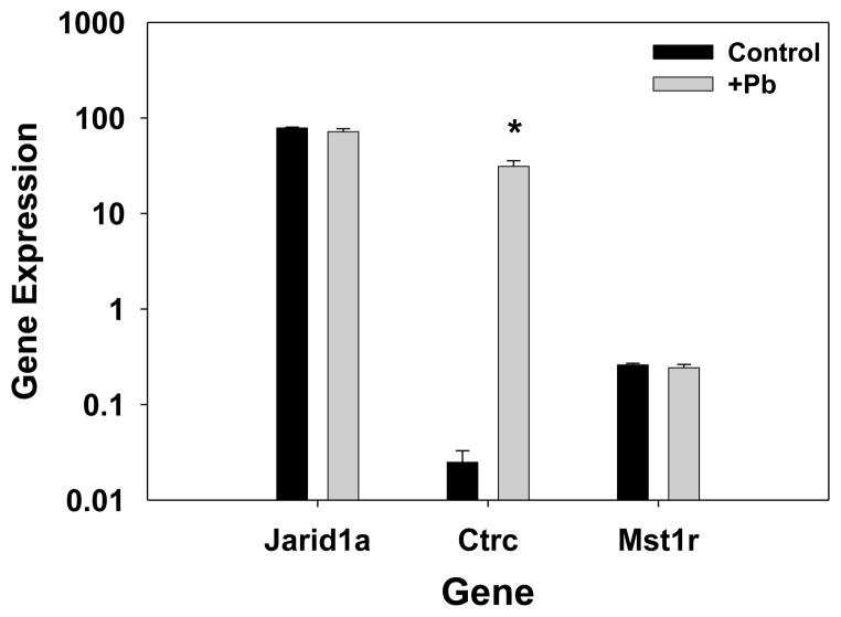
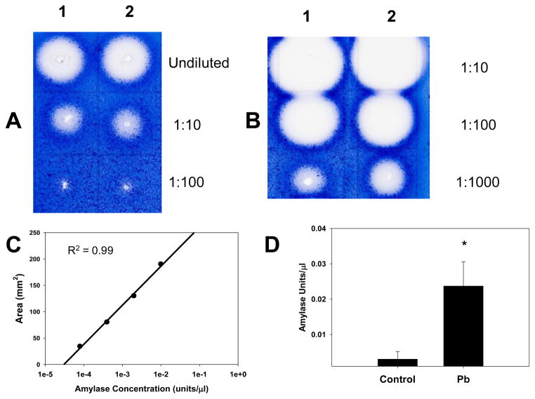
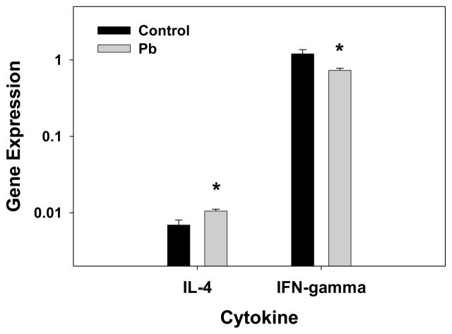
Lead (Pb) is known to alter the functions of numerous organ systems, including the hematopoietic and immune systems. Pb can induce anemia and can lower host resistance to bacterial and viral infections. The anemia is due to Pb's inhibition of hemoglobin synthesis and Pb's induction of membrane changes, leading to early erythrocyte senescence. Pb also increases B-cell activation/proliferation and skews T-cell help (Th) toward Th2 subset generation. The specific mechanisms for many of the Pb effects are, as yet, not completely understood. Therefore, we performed gene expression analysis, via microarray, on RNA from the spleens of developmentally Pb-exposed mice, in order to gain further insight into these Pb effects. Splenic RNA microarray analysis indicated strong up-regulation of genes coding for proteolytic enzymes, lipases, amylase, and RNaseA. The data also showed that Pb affected the expression of many genes associated with innate immunity. Analysis of the microarray results via GeneSifter software indicated that Pb increased apoptosis, B-cell differentiation, and Th2 development. Direct up-regulation by Pb of expression of the gene encoding the heme-regulated inhibitor (HRI) suggested that Pb can decrease erythropoiesis by blocking globin mRNA translation. Pb's high elevation of digestive/catabolizing enzymes could generate immunogenic self peptides. With Pb's potential to induce new self-peptides and to enhance the expression of caspases, cytokines, and other immunomodulators, further evaluation of Pb's involvement in autoimmune phenomena, especially Th2-mediated autoantibody production, and alteration of organ system activities is warranted.
2010 Elsevier Inc. All rights reserved.
Figures
Similar articles
-
Posttranscriptional inhibition of interferon-gamma production by lead.Toxicol Sci. 2007 Mar;96(1):92-100. doi: 10.1093/toxsci/kfl182. Epub 2006 Dec 12. Toxicol Sci. 2007. PMID: 17164472
-
Lead effects on development and function of bone marrow-derived dendritic cells promote Th2 immune responses.Toxicol Appl Pharmacol. 2007 Jul 1;222(1):69-79. doi: 10.1016/j.taap.2007.04.001. Epub 2007 Apr 12. Toxicol Appl Pharmacol. 2007. PMID: 17512567 Free PMC article.
-
Differential effects of lead and cAMP on development and activities of Th1- and Th2-lymphocytes.Toxicol Sci. 1998 Jun;43(2):172-85. doi: 10.1006/toxs.1998.2457. Toxicol Sci. 1998. PMID: 9710959
-
Lead modulation of macrophages causes multiorgan detrimental health effects.J Biochem Mol Toxicol. 2014 Aug;28(8):355-72. doi: 10.1002/jbt.21572. Epub 2014 May 27. J Biochem Mol Toxicol. 2014. PMID: 24863546 Review.
-
STAT4 and STAT6, their role in cellular and humoral immunity and in diverse human diseases.Int Rev Immunol. 2024;43(6):394-418. doi: 10.1080/08830185.2024.2395274. Epub 2024 Aug 26. Int Rev Immunol. 2024. PMID: 39188021 Review.
Cited by
-
Susceptibility to COVID-19 in populations with health disparities: Posited involvement of mitochondrial disorder, socioeconomic stress, and pollutants.J Biochem Mol Toxicol. 2021 Jan;35(1):e22626. doi: 10.1002/jbt.22626. Epub 2020 Sep 9. J Biochem Mol Toxicol. 2021. PMID: 32905655 Free PMC article. Review.
-
Early life environment and developmental immunotoxicity in inflammatory dysfunction and disease.Toxicol Environ Chem. 2011;93(7):1463-1485. doi: 10.1080/02772248.2011.586114. Toxicol Environ Chem. 2011. PMID: 26146439 Free PMC article.
-
Peripheral blood signatures of lead exposure.PLoS One. 2011;6(8):e23043. doi: 10.1371/journal.pone.0023043. Epub 2011 Aug 1. PLoS One. 2011. PMID: 21829687 Free PMC article.
-
Developmental lead effects on behavior and brain gene expression in male and female BALB/cAnNTac mice.Neurotoxicology. 2012 Oct;33(5):1005-20. doi: 10.1016/j.neuro.2012.04.017. Epub 2012 May 16. Neurotoxicology. 2012. PMID: 22609695 Free PMC article.
-
Sub-Chronic Oral Exposure to Iridium (III) Chloride Hydrate in Female Wistar Rats: Distribution and Excretion of the Metal.Dose Response. 2012;10(3):405-14. doi: 10.2203/dose-response.11-052.Iavicoli. Epub 2012 Jan 16. Dose Response. 2012. PMID: 22942873 Free PMC article.
References
-
- Ahamed M, Verma S, Kumar A, Siddiqui MKJ. Delta-aminolevulinic acid dehydratase inhibition and oxidative stress in relation to blood lead among urban adolescents. Human & Exp Toxicol. 2006;25:547–553. - PubMed
-
- Ahamed M, Siddiqui MKJ. Low level lead exposure and oxidative stress: Current opinions. Clinica Chimica Acta. 2007;383:57–64. - PubMed
-
- Anand S, Pal JK. The haem-regulated eukaryotic initiation factor 2alpha kinase: a molecular indicator of lead-toxicity anaemia in rabbits. Biotechnol Appl Biochem. 2002;36:57–62. - PubMed
-
- Arguni E, Arima M, Tsuruoka N, Sakamoto A, Hatano M, Tokuhisa T. JunD/AP-1 and STAT3 are the major enhancer molecules for high Bcl6 expression in germinal center B cells. International Immunology. 2006;18:1079–1089. - PubMed
-
- Bunn TL, Parsons PJ, Kao E, Dietert RR. Exposure to lead during critical windows of embryonic development: Differential immunotoxic outcome based on stage of exposure and gender. Toxicol Sci. 2001;64:57–66. - PubMed
Publication types
MeSH terms
Substances
Grants and funding
LinkOut - more resources
Full Text Sources
