. 2010 Oct 15;53(1):78-84.
doi: 10.1016/j.neuroimage.2010.06.003. Epub 2010 Jun 11.

Cost function masking during normalization of brains with focal lesions: still a necessity?

Affiliations

Cost function masking during normalization of brains with focal lesions: still a necessity?

Sarah M Andersen et al. Neuroimage. .

Abstract

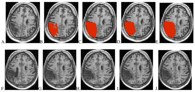
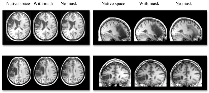
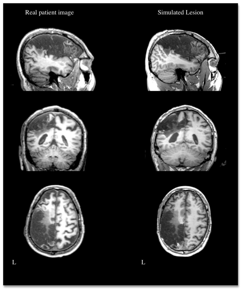
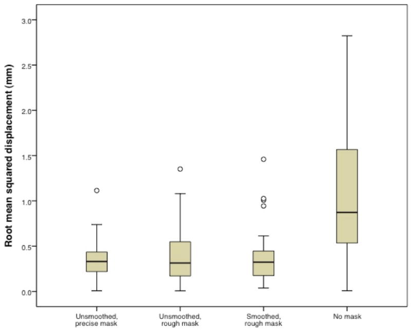
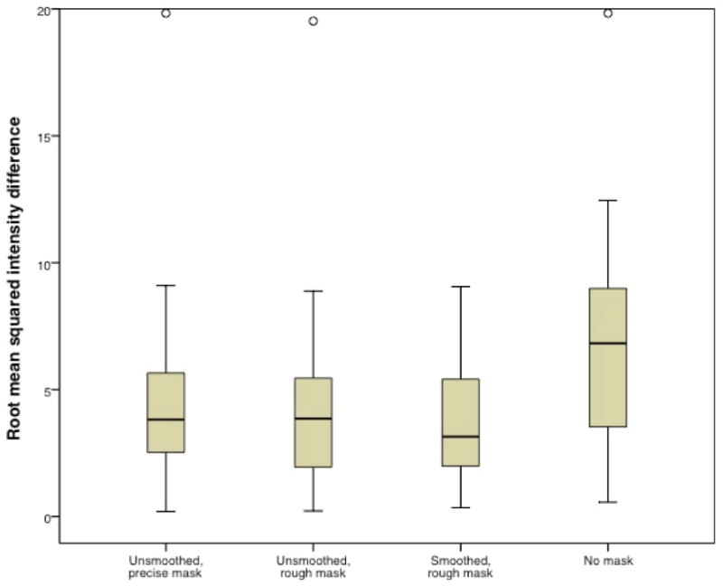
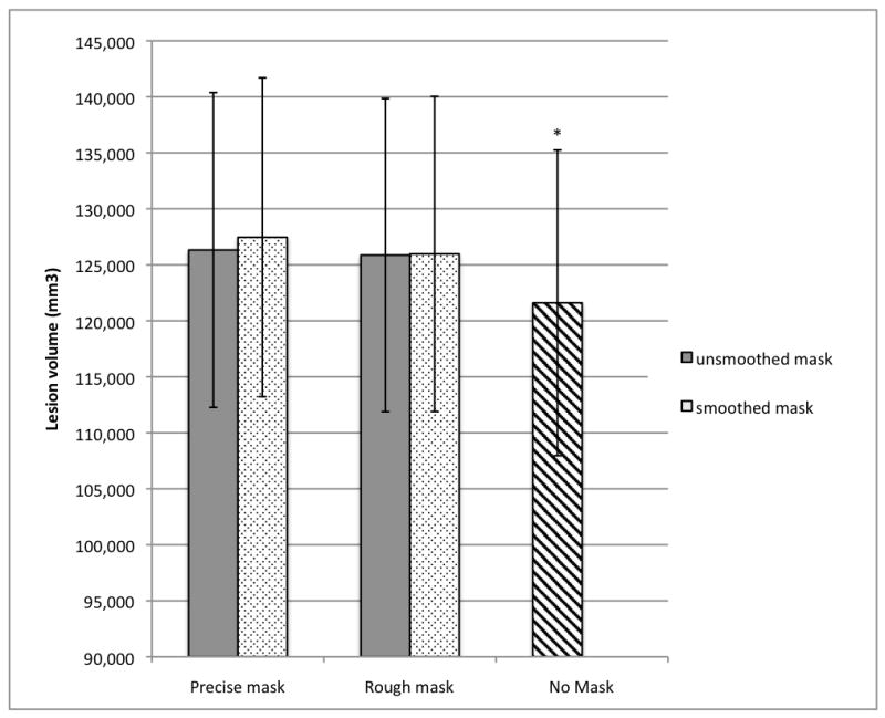
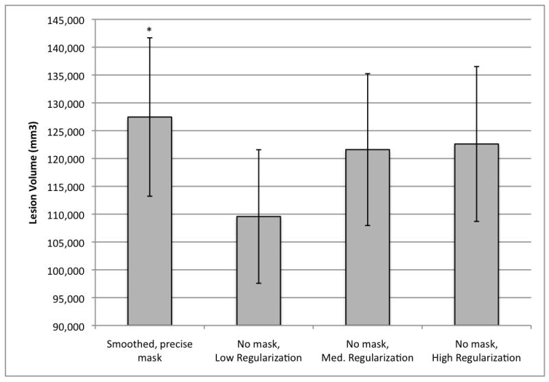
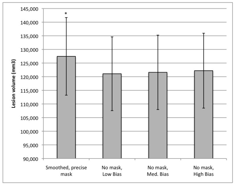
Although normalization of brain images is critical to the analysis of structural damage across individuals, loss of tissue due to focal lesions presents challenges to the available normalization algorithms. Until recently, cost function masking, as advocated by Brett and colleagues (2001), was the accepted method to overcome difficulties encountered when normalizing damaged brains; however, development of the unified segmentation approach for normalization in SPM5 (Ashburner & Friston, 2005) offered an alternative. Crinion et al. (2007) demonstrated this approach produced normalization results without cost function masking that appeared to be robust to lesion effects when tested using the same simulated lesions studied by Brett et al. (2001). The present study sought to confirm the validity of this approach in brains with focal damage due to vascular events. To do so, we examined outcomes of normalization using unified segmentation with and without cost function masking in 49 brain images with chronic stroke. Lesion masks were created using two approaches (precise and rough drawings of lesion boundaries), and normalization was implemented with both smoothed and unsmoothed versions of the masks. We found that failure to employ cost function masking produced less accurate results in real and simulated lesions, compared to masked normalization, both in terms of deformation field displacement and voxelwise intensity differences. Additionally, unmasked normalization led to significant underestimation of lesion volume relative to all four masking conditions, especially in patients with large lesions. Taken together, these findings suggest cost function masking is still necessary when normalizing brain images with chronic infarcts.


Figures

Figure 1
Figure 1
Methods of lesion masking in approximately aligned axial slices from one MCA stroke patient. Upper images (A-E): lesion in native space and shown with four masking conditions. Lower images (F-J): results after spatial normalization. A. Native space lesion without mask B. Native space brain with unsmoothed, precise lesion mask C. Native space brain with smoothed, precise lesion mask D. Native space brain with unsmoothed, rough lesion mask E. Native space brain with smoothed, rough lesion mask F. Normalized brain using no lesion mask G. Normalized brain using unsmoothed, precise lesion mask H. Normalized brain using smoothed, precise lesion mask I. Normalized brain using unsmoothed, rough lesion mask J. Normalized brain using smoothed, rough lesion mask
Figure 2
Figure 2
Examples of four different brains with left hemisphere lesions in native space and normalization results accomplished with and without cost function masking (using a smoothed, precise mask).
Figure 3
Figure 3
Example of a real brain lesion and a simulated lesion generated by inserting the region of damage in a healthy brain. L = left.
Figure 4
Figure 4
Root mean squared displacement values comparing outcomes using the smoothed, precise mask relative to the other masking conditions. Open circle indicates outlier. Root mean squared difference for the unmasked condition (mean = 1.07, s.d. = 0.72) is significantly larger than for each of the other conditions (unsmoothed, precise mask mean = 0.347, s.d. = 0.203; unsmoothed, rough mask mean = 0.403, s.d. = 0.311; smoothed, rough mask mean = 0.370, s.d. = 0.275).
Figure 5
Figure 5
Root mean squared intensity difference comparing outcomes using the smoothed, precise mask relative to the other masking conditions. Open circle indicates outlier. Root mean squared difference for the unmasked condition (mean = 6.3, s.d. = 3.8) is significantly larger than for each of the other conditions (unsmoothed, precise mask mean = 4.3, s.d. = 3.2; unsmoothed, rough mask mean = 4.0, s.d. = 3.2; smoothed, rough mask mean = 3.8, s.d. = 2.2).
Figure 6
Figure 6
Mean lesion volumes obtained with the different normalization methods (n = 49). *Unmasked lesion volume (121,602 mm3, S.E. = 13,646) significantly smaller than lesion volumes obtained in all masked conditions (unsmoothed, precisely masked lesion volume = 126,322 mm3, S.E. = 14,056; smoothed, precisely masked lesion volume = 127,458 mm3, S.E. = 14,232; unsmoothed, roughly masked lesion volume = 125,863 mm3, S.E. = 13,975; smoothed, roughly masked lesion volume = 125,967 mm3, S.E. = 14,069).
Figure 7
Figure 7
Mean lesion volumes obtained with variations of the warping regularization parameter in the unmasked condition (n=49). Bias regularization was held constant at 0.0001 for all conditions. * Smoothed, precisely masked lesion volume (127,458 mm3, S.E. = 14,232) significantly larger than lesion volumes obtained in all unmasked conditions (low regularization = 109,580 mm3, S.E. = 11,999; medium regularization = 121,602 mm3, S.E. = 13,646; and high regularization = 122,611 mm3, S.E. = 13,911).
Figure 8
Figure 8
Mean lesion volumes obtained with variations of the bias regularization parameter in the unmasked condition (n=49). Warping regularization was held at medium for all conditions. Low bias = 0.00001; medium bias = 0.0001; high bias = 0.001. *Smoothed, precisely masked lesion volume (127,458 mm3, S.E. = 14,232) significantly larger than lesion volumes obtained in all unmasked conditions (low bias = 121,079 mm3, S.E. = 13,519; medium bias = 121,602 mm3, S.E. = 13,646; and high bias = 122,211 mm3, S.E. = 13,731).

References

    1. Ashburner J, Friston KJ. Unified segmentation. NeuroImage. 2005;26:839–851. - PubMed
    1. Brett M, Leff AP, Rorden C, Ashburner J. Spatial normalization of brain images with focal lesions using cost function masking. NeuroImage. 2001;14(2):486–500. - PubMed
    1. Crinion J, Ashburner J, Leff AP, Brett M, Price C, Friston K. Spatial normalization of lesioned brains: Performance evaluation and impact on fMRI analyses. NeuroImage. 2007;37(3):866–875. - PMC - PubMed
    1. Kim J, Avants B, Patel S, Whyte J. Spatial normalization of injured brains for neuroimaging research: An illustrative introduction of available options. 2007. http://www.ncrrn.org/papers/methodology_papers/sp_norm_kim.pdf.
    1. Klein A, Andersson J, Ardekani BA, Ashburner J, Avants B, Chiang M-C, Christensen GE, Collins DL, Gee J, Hellier P, Song JH, Jenkinson M, Lepage C, Rueckert D, Thompson P, Vercauteren T, Woods RP, Mann JJ, Parsey RV. Evaluation of 14 nonlinear deformation algorithms applied to human brain MRI registration. NeuroImage. 2009;46:786–802. - PMC - PubMed

Publication types