. 2010 Jun 25;7(6):463-73.
doi: 10.1016/j.chom.2010.05.012.

Human formyl peptide receptor 2 senses highly pathogenic Staphylococcus aureus

Affiliations

Human formyl peptide receptor 2 senses highly pathogenic Staphylococcus aureus

Dorothee Kretschmer et al. Cell Host Microbe. .

Abstract

Virulence of emerging community-associated methicillin-resistant Staphylococcus aureus (CA-MRSA) and other highly pathogenic S. aureus strains depends on their production of phenol-soluble modulin (PSM) peptide toxins, which combine the capacities to attract and lyse neutrophils. The molecular basis of PSM-stimulated neutrophil recruitment has remained unclear. Here, we demonstrate that the human formyl peptide receptor 2 (FPR2/ALX), which has previously been implicated in control of endogenous inflammatory processes, senses PSMs at nanomolar concentrations and initiates proinflammatory neutrophil responses to CA-MRSA. Specific blocking of FPR2/ALX or deletion of PSM genes in CA-MRSA severely diminished neutrophil detection of CA-MRSA. Furthermore, a specific inhibitor of FPR2/ALX and of its functional mouse counterpart blocked PSM-mediated leukocyte infiltration in vivo in a mouse model. Thus, the innate immune system uses a distinct FPR2/ALX-dependent mechanism to specifically sense bacterial peptide toxins and detect highly virulent bacterial pathogens. FPR2/ALX represents an attractive target for new anti-infective or anti-inflammatory strategies.


Figures

Figure 1
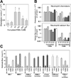
Figure 1. Formylated and Non-Formylated PSM Peptides Elicit Chemotaxis and Calcium Ion Fluxes in Human Neutrophils
(A) PSMα3 stimulates chemotaxis in human neutrophils at nanomolar concentrations with a typical bell-shaped dose/response curve. (B) Both, formylated and non-formylated core-genome encoded PSMs (PSMα1–4, PSMβ1–2, δ-toxin) and PSM-mec induce chemotaxis and chemotaxis-associated calcium fluxes in human neutrophils (values normalized to 1 µM) (C) Neutrophil stimulation by PSMs is sensitive to pertussis toxin and the FPR2/ALX-specific inhibitor FLIPr but not the FPR1-specific inhibitor CHIPS. fMLF and MMK1 are synthetic control ligands of FPR1 and FPR2/ALX, respectively. Data represent means ± SEM of at least three independent experiments. *, P < 0.05; **, P < 0.005; ***, P < 0.001 versus lowest peptide concentration (A), or no inhibition (C). All values were significant versus buffer control in (B) (P < 0.05 or lower). δ, δ-toxin; fl, fluorescence.
Figure 2
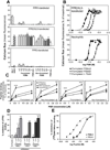
Figure 2. PSMs Specifically Activate and Bind to FPR2/ALX
(A) PSMs stimulate profound calcium fluxes in FPR2/ALX-transfected but not in FPR1 or FPR3-transfected cells. Untransfected HL60 cells exhibited no significant responses (mean fluorescence values below 1, data not shown). fMLF and MMK1 are synthetic control ligands of FPR1 and FPR2/ALX, respectively. (B) Dose-response curves for Ca2+ fluxes induced by formylated PSMα3, β2, and δ-toxin in FPR2/ALX-transfected HL60 or neutrophils. (C) 5-carboxytetramethylrhodamin-labeled PSMs bind specifically to FPR2/ALX-transfected HL60 cells. Maximum detected binding to FPR2/ALX-transfected cells was defined as 100%. (D) PSMs prevent binding of a FPR2/ALX-specific phycoerythrin-labeled monoclonal antibody (αFPR2) from FPR2/ALX-transfected HL60 cells in a dose-dependent manner. Fluorescence of samples with the phycoerythrin-labeled Isotypecontrol was defined as the maximally achievable inhibition of binding (100% inhibition). (E) PSMα3 displaces the synthetic 125I-labeld FPR2/ALX ligand WKYMVm from transiently FPR2/ALX-transfected cells. Unlabeled FPR2/ALX ligand CKβ8-1 was used as a positive control Data represent mean ± SEM of at least three independent experiments. *, P < 0.05; ***, P < 0.001 versus untransfected cells (A, C) or versus no peptide (D). δ, δ-toxin.
Figure 3
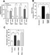
Figure 3. In Vitro and In Vivo Inhibition of PSM-Mediated Neutrophil Activation and Mobilization in Mice by WRW4
(A) WRW4 blocks calcium fluxes in response to MMK1 and PSMs but not to fMLF in mouse neutrophils. (B) Intraperitoneal challenge of mice with S. aureus USA300 induces granulocyte infiltration, which can be blocked by WRW4. Granulocytes were identified by Gr1+ staining. (C) PSMα2 leads to leukocyte infiltration into mouse air pouches and this process can be blocked by WRW4 but not by its scrambled congener wwrw3. Data represent means ± SEM of at least three (A), 5–8 independent experiments with the same number of mice per group (B), or four independent experiments with 2–3 mice per group (C). *, P < 0.05 **, P < 0.005; ***, P < 0.001; ns, not significantly different as calculated by Student’s t-test (A, B) or Mann-Whitney u-test (C).
Figure 4
Figure 4. FPR2/ALX Initiates Exuberant Proinflammatory Neutrophil Responses to PSMs in CA-MRSA Culture Supernatants
Calcium flux stimulated by CA-MRSA culture supernatants is FPR2/ALX-dependent in neutrophils (A) and receptor-transfected HL60 cells (B). Untransfected HL60 cells exhibited no significant responses (mean fluorescence values below 1, data not shown). (C) FPR2/ALX-transfected HL60 cells respond exuberantly to CA-MRSA (USA300, USA400) but only moderately to HA-MRSA (COL, Mu50, N315) while FPR1-mediated responses differ only slightly between CA-MRSA and HA-MRSA. (D) FLIPr inhibits USA300-stimulated neutrophil chemotaxis and IL-8 release. The indicated S. aureus wild-type (WT) or PSM gene deletion mutants (Δα, Δβ, or Δδ) were used in A–D. USA400 supernatants yielded similar results as shown for USA300 in (A, B, D) (data not shown). Data represent means ± SEM of at least three independent experiments. **, P < 0.005; ***, P < 0.001 versus no inhibition (A, D) or untransfected HL60 (B).
Figure 5
Figure 5. Proposed Role of FPR2/ALX-Mediated Sensing of PSM Peptide Toxins
FPR2/ALX allows neutrophils to distinguish between S. aureus with low and high virulence potential by sensing the concentration of PSMs and causes neutrophil infiltration in response to highly pathogenic S. aureus. In contrast, FPR1 mediates moderate neutrophil activation in response to S. aureus irrespective of virulence. CHIPS and FLIPr are thought to prevent neutrophil influx during certain stages of human colonization and infection when it is most critical for the bacteria to prevent recruitment of leukocytes (de Haas et al., 2004; Prat et al., 2006).

Comment in

References

    1. Alvarez V, Coto E, Setien F, Gonzalez-Roces S, Lopez-Larrea C. Molecular evolution of the N-formyl peptide and C5a receptors in non-human primates. Immunogenetics. 1996;44:446–452. - PubMed
    1. Bubeck WJ, Bae T, Otto M, DeLeo FR, Schneewind O. Poring over pores: alpha-hemolysin and Panton-Valentine leukocidin in Staphylococcus aureus pneumonia. Nat. Med. 2007;13:1405–1406. - PubMed
    1. Chambers HF, DeLeo FR. Waves of resistance:Staphylococcus aureus in the antibiotic era. Nat. Rev. Microbiol. 2009;7:629–641. - PMC - PubMed
    1. Christophe T, Karlsson A, Dugave C, Rabiet MJ, Boulay F, Dahlgren C. The synthetic peptide Trp-Lys-Tyr-Met-Val-Met-NH2 specifically activates neutrophils through FPRL1/lipoxin A4 receptors and is an agonist for the orphan monocyte-expressed chemoattractant receptor FPRL2. J. Biol. Chem. 2001;276:21585–21593. - PubMed
    1. Cui Y, Le Y, Yazawa H, Gong W, Wang JM. Potential role of the formyl peptide receptor-like 1 (FPRL1) in inflammatory aspects of Alzheimer's disease. J. Leukoc. Biol. 2002;72:628–635. - PubMed

Publication types

MeSH terms