. 2010 Nov 1;88(2):344-51.
doi: 10.1093/cvr/cvq184. Epub 2010 Jun 11.

Sphingosine-1-phosphate modulation of basal permeability and acute inflammatory responses in rat venular microvessels

Affiliations

Sphingosine-1-phosphate modulation of basal permeability and acute inflammatory responses in rat venular microvessels

Roger H Adamson et al. Cardiovasc Res. .

Abstract

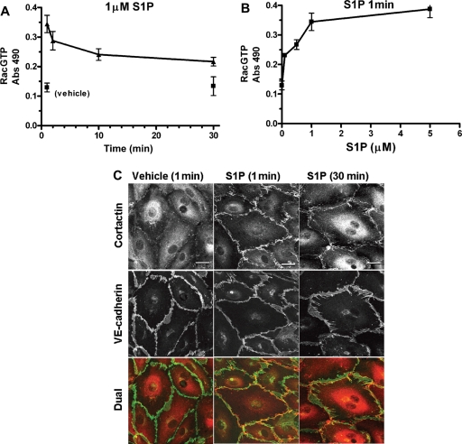
Aims: Although several cultured endothelial cell studies indicate that sphingosine-1-phosphate (S1P), via GTPase Rac1 activation, enhances endothelial barriers, very few in situ studies have been published. We aimed to further investigate the mechanisms whereby S1P modulates both baseline and increased permeability in intact microvessels.

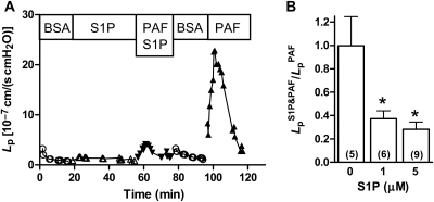
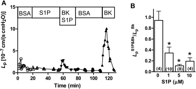
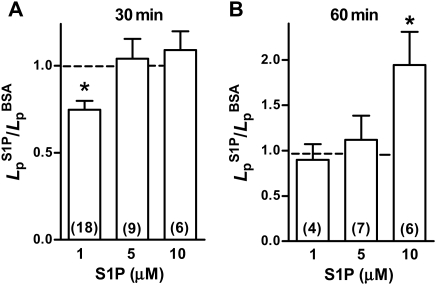
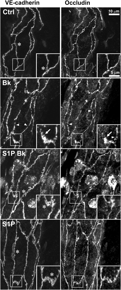
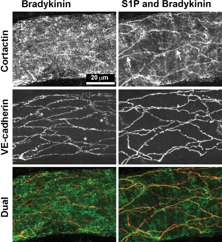
Methods and results: We measured attenuation by S1P of platelet-activating factor (PAF)- or bradykinin (Bk)-induced hydraulic conductivity (L(p)) increase in mesenteric microvessels of anaesthetized rats. S1P alone (1-5 µM) attenuated by 70% the acute L(p) increase due to PAF or Bk. Immunofluorescence methods in the same vessels under identical experimental conditions showed that Bk or PAF stimulated the loss of peripheral endothelial cortactin and rearrangement of VE-cadherin and occludin. Our results are the first to show in intact vessels that S1P pre-treatment inhibited rearrangement of VE-cadherin and occludin induced by PAF or Bk and preserved peripheral cortactin. S1P (1-5 µM, 30 min) did not increase baseline L(p). However, 10 µM S1P (60 min) increased L(p) two-fold.

Conclusion: Our results conform to the hypothesis that S1P inhibits acute permeability increase in association with enhanced stabilization of peripheral endothelial adhesion proteins. These results support the idea that S1P can be useful to attenuate inflammation by enhancing endothelial adhesion through activation of Rac-dependent pathways.


Figures

Figure 1
Figure 1
(A) S1P inhibits acute Lp response to PAF. Representative data from one experiment show that treatment with S1P (1 µM) inhibits the first test with PAF (10 nM), whereas a second test in the absence of S1P after recovery to baseline shows a typical PAF response. (B) Dose–response relation for inhibition of PAF by S1P. The ratio of the peak Lp response to PAF (10 nM) in the presence of S1P to the peak value in the absence of S1P is shown. Both 1 and 5 µM S1P inhibit about 65% of the acute Lp peak response. Control value shows that repeated treatment with PAF yields comparable peaks (*different from control, P < 0.05, Wilcoxon's signed-rank test, n for each group shown in parentheses).
Figure 2
Figure 2
(A) S1P inhibits acute Lp response to Bk. Using the same experimental design as in Figure 1 for PAF experiments, S1P (1 µM) is shown to partially inhibit the acute peak Lp response to Bk (10 nM) during the first Bk test Lp these data from a representative vessel. After recovery to baseline, the second Bk test responds with a typical rapid increase and recovery. (B) Dose–response relation for S1P inhibition of Bk. S1P at 1, 5, and 10 µM strongly reduces the peak Lp response to Bk (10 nM), but even at the highest concentration it does not fully block the response. Repeated tests in the absence of S1P show that multiple Bk tests yield similar peaks (*different from control, P < 0.05, Wilcoxon's signed-rank test, n for each group shown in parentheses).
Figure 3
Figure 3
Effect of S1P on baseline Lp. (A) The ratio of Lp measured after 30 min of S1P relative to the value measured with vehicle solution alone is shown. Only at 1µM is the Lp significantly reduced to about 70% of control. At higher concentrations, there was no significant difference from control (*different from value of 1; P < 0.05, Wilcoxon's signed-rank test, n for each group shown in parentheses). (B) The ratio of Lp measured after 60 min of S1P relative to the Lp in the absence of S1P is shown. With 1 or 5 µM, there was no change from control. At 10 µM, S1P Lp increased by about two-fold (*different from value of 1; P < 0.05, Wilcoxon's signed-rank test, n for each group shown in parentheses).
Figure 4
Figure 4
S1P prevents rearrangement of VE-cadherin and occludin in vivo. Segment of a vessel wall dual labelled for VE-cadherin and occludin from a vessel perfused with vehicle solution (Ctrl) shows continuous label for both proteins. (The images are projections from stacks of images taken of three-dimensional vessel wall so that some junctions appear to be incomplete where they pass out of the collection volume.) A representative vessel that was perfused with Bk (10 nM, 5 min) shows discrete gaps (arrowheads) in both VE-cadherin and occludin, as well as spikes oriented perpendicularly to the junction (arrows). The Lp of the Bk-treated vessel was 14 × 10−7 cm/(s cmH2O) immediately before fixation (Lp values of Ctrl, S1P-Bk, and S1P vessels were 1.2, 1.4, and 0.8, respectively, i.e. they were low and normal). In vessels pre-treated with S1P (5 µM, 30 min) prior to Bk, there were no prominent gaps or rearrangements of the junction protein patterns (S1P-Bk). Vessels treated with S1P alone (5 µM, 30 min) looked very similar to vehicle controls with continuous junctions and exhibited broad areas of VE-cadherin (asterisks).
Figure 5
Figure 5
Effects of S1P on cultured HMVEC. (A) Rac activation, shown over time when exposed to S1P (1 µM), rapidly rises relative to vehicle treatment and remains elevated for at least 30 min (mean ± SEM). (B) Dose–response relationship for Rac activation in the presence of S1P (1 min) shows that level of activation at 1 µM is nearly equal to that at 5 µM. (C) Representative cultures of HMVEC double labelled for cortactin and VE-cadherin show that treatment with 1 µM S1P induces strong peripheral cortactin at both 1 and 30 min relative to vehicle control. VE-cadherin after S1P treatment was not different from fully confluent, quiescent monolayers. Scale bar 20 µm.
Figure 6
Figure 6
S1P localizes cortactin to periphery in vivo. Representative vessels, perfused with Bk (10 nM) and pre-treated with S1P (5 µM, 30 min) prior to Bk, respectively, were dual labelled for cortactin and VE-cadherin. The Bk vessel [final Lp was 34 × 10−7 cm/(scmH2O)] shows distinctive gaps and lateral spikes (asterisks) in VE-cadherin, whereas the vessel pre-treated with S1P prior to Bk [final Lp was 2 × 10⁻⁷ cm/(scmH2O)] had continuous VE-cadherin. Cortactin was diffuse in the vessel wall of the Bk vessel, but in the S1P pre-treated vessel, cortactin was clearly enhanced at the periphery of many endothelial cells (arrows) where it is seen near the VE-cadherin in the dual label image. (When these specimens were mounted, the vessels collapsed and therefore the labelled endothelial junctions from opposite sides of the vessel appear to cross one another.)

References

    1. Adamson RH, Zeng M, Adamson GN, Lenz JF, Curry FE. PAF- and bradykinin-induced hyperpermeability of rat venules is independent of actin-myosin contraction. Am J Physiol Heart Circ Physiol. 2003;285:H406–417. - PubMed
    1. Garcia JG, Davis HW, Patterson CE. Regulation of endothelial cell gap formation and barrier dysfunction: role of myosin light chain phosphorylation. J Cell Physiol. 1995;163:510–522. doi:10.1002/jcp.1041630311. - DOI - PubMed
    1. van Nieuw Amerongen GP, van Hinsbergh VW. Targets for pharmacological intervention of endothelial hyperpermeability and barrier function. Vascul Pharmacol. 2002;39:257–272. doi:10.1016/S1537-1891(03)00014-4. - DOI - PubMed
    1. McVerry BJ, Garcia JG. In vitro and in vivo modulation of vascular barrier integrity by sphingosine 1-phosphate: mechanistic insights. Cell Signal. 2005;17:131–139. doi:10.1016/j.cellsig.2004.08.006. - DOI - PubMed
    1. Garcia JG, Liu F, Verin AD, Birukova A, Dechert MA, Gerthoffer WT, et al. Sphingosine 1-phosphate promotes endothelial cell barrier integrity by Edg-dependent cytoskeletal rearrangement. J Clin Invest. 2001;108:689–701. - PMC - PubMed

Publication types

MeSH terms