. 2010 Oct;38(19):6404-17.
doi: 10.1093/nar/gkq504. Epub 2010 Jun 11.

Binding site number variation and high-affinity binding consensus of Myb-SANT-like transcription factor Adf-1 in Drosophilidae

Affiliations

Binding site number variation and high-affinity binding consensus of Myb-SANT-like transcription factor Adf-1 in Drosophilidae

Michael Lang et al. Nucleic Acids Res. 2010 Oct.

Abstract

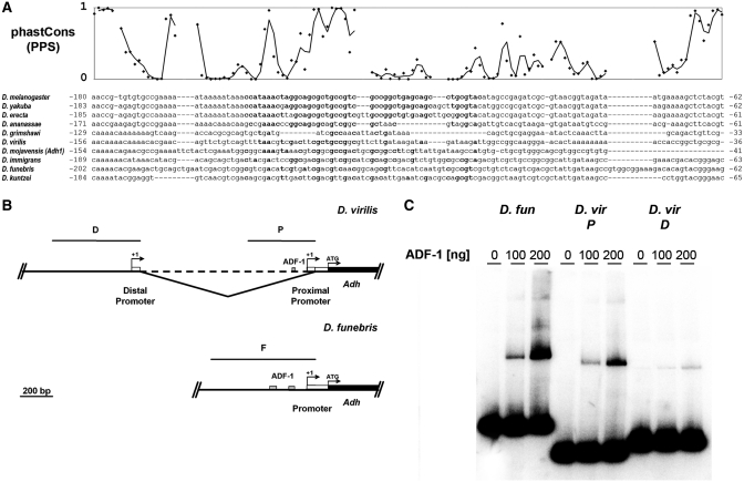
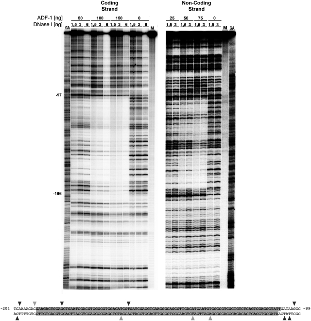
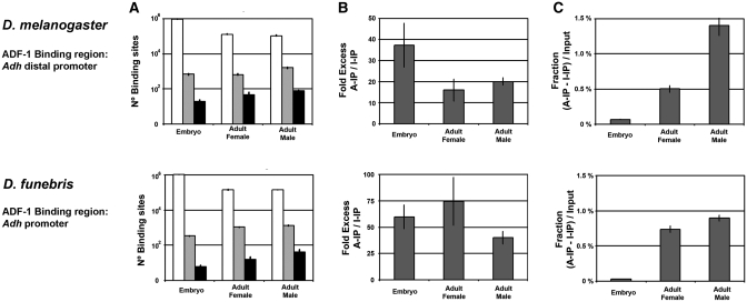
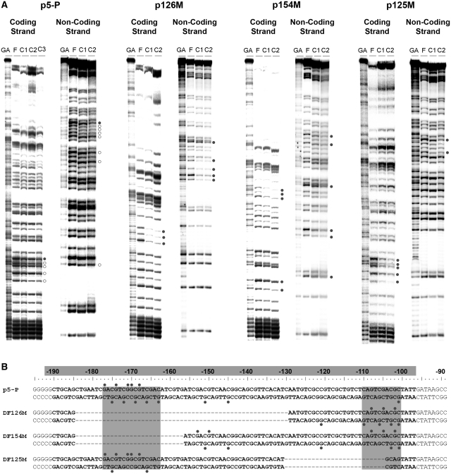
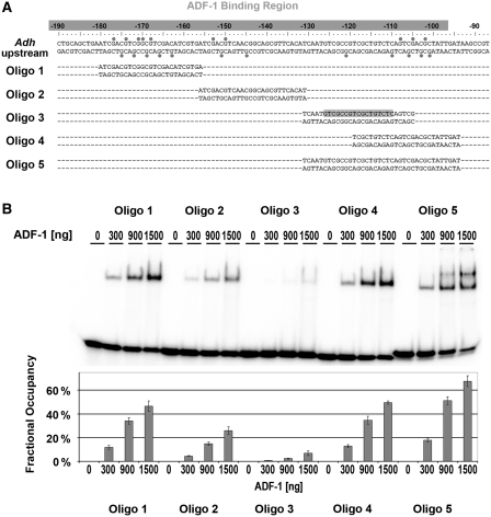
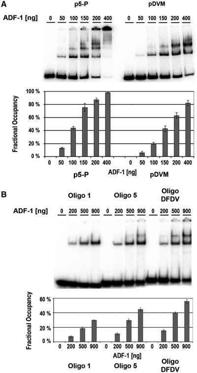
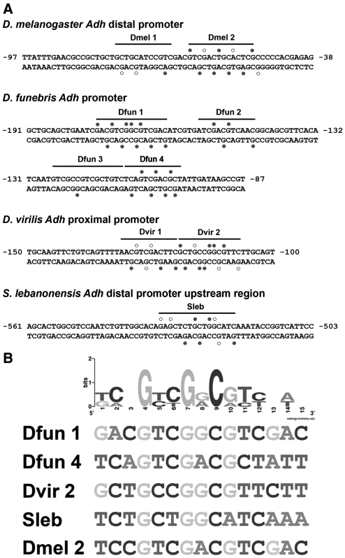
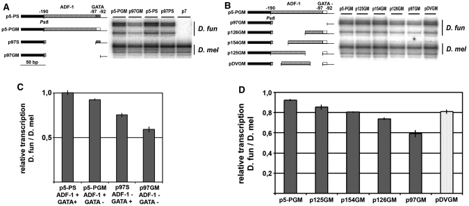
There is a growing interest in the evolution of transcription factor binding sites and corresponding functional change of transcriptional regulation. In this context, we have examined the structural changes of the ADF-1 binding sites at the Adh promoters of Drosophila funebris and D. virilis. We detected an expanded footprinted region in D. funebris that contains various adjacent binding sites with different binding affinities. ADF-1 was described to direct sequence-specific DNA binding to sites consisting of the multiple trinucleotide repeat . The ADF-1 recognition sites with high binding affinity differ from this trinucleotide repeat consensus sequence and a new consensus sequence is proposed for the high-affinity ADF-1 binding sites. In vitro transcription experiments with the D. funebris and D. virilis ADF-1 binding regions revealed that stronger ADF-1 binding to the expanded D. funebris ADF-1 binding region only moderately lead to increased transcriptional activity of the Adh gene. The potential of this regional expansion is discussed in the context of different ADF-1 cellular concentrations and maintenance of the ADF-1 stimulus. Altogether, evolutionary change of ADF-1 binding regions involves both, rearrangements of complex binding site cluster and also nucleotide substitutions within sites that lead to different binding affinities.


Figures

Figure 1.
Figure 1.
ADF-1 binding sites at the Adh proximal promoter. (A) Conservation evaluation with phastCons (61). Conservation estimates are indicated by diamonds, the line shows the moving average. (B) Putative ADF-1 binding sites in the Adh promoter regions of D. virilis (43) and D. funebris (32), indicated by gray boxes (C) EMSA with corresponding promoter regions, using S. lebanonensis ADF-1. P: D. virilis proximal promoter, D: D. virilis distal promoter, D. vir: D. virilis, D. fun: D. funebris.
Figure 2.
Figure 2.
DNase I protection of the ADF-1 Binding region at the D. funebris Adh promoter. This experiment was performed with the ADF-1 protein of D. funebris. M: Mock experiment, GA: Maxam Gilbert G+A sequencing ladder. Hypersensitive sites are indicated by black triangles, weak hypersensitivity in gray (<150% at 50 ng ADF-1 and 3 ng DNase I).
Figure 3.
Figure 3.
In vivo binding of ADF-1 at the D. funebris Adh promoter region. (A) Quantification of ADF-1 binding sites. The input sample is indicated in white, the anti-ADF-1 ChIP in gray and the control ChIP in black. (B) Enrichment of ADF-1 binding sites in anti-ADF-1 ChIP relative to the control IgG ChIP experiments. (C) The total fraction of specifically immunoprecipitated DNA with respect to the input sample. A-IP, anti-ADF-1 ChIP; I-IP, control ChIP experiment with unspecific IgG.
Figure 4.
Figure 4.
ADF-1 contacts to two subregions of the DNaseI footprint at the D. funebris Adh promoter. (A) Methylation interference with D. funebris ADF-1 and the partial deletion constructs, closed circles indicate guanine residues that interfere with ADF-1 binding when methylated, weak interference sites are indicated by open circles. F: sequencing ladder from unbound DNA, C1, C2 and C3: sequencing ladder from bound DNA, the numbers indicate the order of supershifts. (B) ADF-1 DNase I footprint is indicated by the gray bar, subregions with abundant methylation interference sites are shaded gray.
Figure 5.
Figure 5.
ADF-1 binding affinity for the different elements in the binding region. (A) Double-stranded oligonucleotides. The ADF-1 footprint is indicated by a gray bar. Methylation interference sites are indicated by gray closed circles and the sequence that follows the England’s ADF-1 binding consensus is highlighted in the forward strand of Oligo 3. (B) Comparison of ADF-1 binding affinity to the Oligonucleotides 1–5. Representative EMSA’s are shown with D. funebris ADF-1.
Figure 6.
Figure 6.
Comparison of ADF-1 binding affinity to the D. virilis Adh proximal promoter and D. funebris promoter. (A) Binding of ADF-1 of D. funebris to the entire binding regions of D. funebris and D. virilis. (B) Binding of ADF-1 to the double-stranded oligonucleotides DFDV, Oligos 1 and 5.
Figure 7.
Figure 7.
ADF-1 binding sites upstream of the Adh gene in Drosophilidae and ADF-1 high-affinity binding consensus. (A) ADF-1 binding sites are recognized as regions of maximal 17 bp that contain at least three nearby methylation interference sites. The site Dfun 3, which contains the England’s consensus, shows the lowest binding affinity by ADF-1 as determined by the binding to Oligos 5 and 3. The closed circles indicate strong methylation interference while open circles indicate partial methylation interference. Sequence positions indicate the distances to the transcriptional start site. (B) The sequence logo was established with Weblogo (www.weblogo.berkely.edu).
Figure 8.
Figure 8.
In vitro transcription of D. funebris Adh deletion constructs. (A) Deletion mutation constructs, the intact GATA element is indicated by the gray box and the mutated element by the white box; the ADF-1 binding region is marked with the dashed boxes. Primer extension bands are labeled D. fun, D. funebris transcripts; D. mel, D. melanogaster transcripts. (B) Quantification of primer extension band intensities. Four distinct homogenous groups identified by multiple range test (95% CI). (C) Partial deletion constructs of the ADF-1 binding region at the D. funebris Adh promoter. (D) Quantification of primer extension band intensities. Five distinct homogenous groups identified by multiple range test (95% CI), joining pDVGM and p154GM.

Similar articles

Cited by

References

    1. King M-C, Wilson AC. Evolution at two levels in humans and chimpanzees. Science. 1975;188:107–116. - PubMed
    1. Carroll SB. Evo-devo and an expanding evolutionary synthesis: a genetic theory of morphological evolution. Cell. 2008;134:25–36. - PubMed
    1. Prud'homme B, Gompel N, Carroll SB. Emerging principles of regulatory evolution. Proc. Natl Acad. Sci. USA. 2007;104:8605–8612. - PMC - PubMed
    1. Ludwig MZ. Functional evolution of noncoding DNA. Curr. Opin. Genet. Dev. 2002;12:634–639. - PubMed
    1. Ludwig MZ, Palsson A, Alekseeva E, Bergman CM, Nathan J, Kreitman M. Functional Evolution of a cis-Regulatory Module. PLoS Biol. 2005;3:e93. - PMC - PubMed

Publication types