. 2010 Sep;192(17):4267-74.
doi: 10.1128/JB.00403-10. Epub 2010 Jun 11.

Phosphorylation and dephosphorylation among Dif chemosensory proteins essential for exopolysaccharide regulation in Myxococcus xanthus

Affiliations

Phosphorylation and dephosphorylation among Dif chemosensory proteins essential for exopolysaccharide regulation in Myxococcus xanthus

Wesley P Black et al. J Bacteriol. 2010 Sep.

Abstract

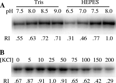
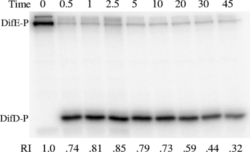
Myxococcus xanthus social gliding motility, which is powered by type IV pili, requires the presence of exopolysaccharides (EPS) on the cell surface. The Dif chemosensory system is essential for the regulation of EPS production. It was demonstrated previously that DifA (methyl-accepting chemotaxis protein [MCP]-like), DifC (CheW-like), and DifE (CheA-like) stimulate whereas DifD (CheY-like) and DifG (CheC-like) inhibit EPS production. DifD was found not to function downstream of DifE in EPS regulation, as a difD difE double mutant phenocopied the difE single mutant. It has been proposed that DifA, DifC, and DifE form a ternary signaling complex that positively regulates EPS production through the kinase activity of DifE. DifD was proposed as a phosphate sink of phosphorylated DifE (DifE approximately P), while DifG would augment the function of DifD as a phosphatase of phosphorylated DifD (DifD approximately P). Here we report in vitro phosphorylation studies with all the Dif chemosensory proteins that were expressed and purified from Escherichia coli. DifE was demonstrated to be an autokinase. Consistent with the formation of a DifA-DifC-DifE complex, DifA and DifC together, but not individually, were found to influence DifE autophosphorylation. DifD, which did not inhibit DifE autophosphorylation directly, was found to accept phosphate from autophosphorylated DifE. While DifD approximately P has an unusually long half-life for dephosphorylation in vitro, DifG efficiently dephosphorylated DifD approximately P as a phosphatase. These results support a model where DifE complexes with DifA and DifC to regulate EPS production through phosphorylation of a downstream target, while DifD and DifG function synergistically to divert phosphates away from DifE approximately P.


Figures

FIG. 1.
FIG. 1.
Fractionation of DifE by Ni-affinity chromatography. Equal volumes of selected fractions from Ni-affinity chromatography were subjected to SDS-PAGE and Coomassie blue staining (A) or autophosphorylation assays (B). The fraction numbers indicated at the top of the figure apply to both panels (fractions 4 to 10). The positions of DifE and DifE∼P are indicated on the left. For the phosphorylation studies in panel B, the radioactivity associated with 32P-labeled DifE was analyzed by phosphorimaging as described in Materials and Methods. Indicated at the bottom of the figure is the relative intensity (RI) of each band, which was normalized to the one with the highest radioactivity.
FIG. 2.
FIG. 2.
Optimization of conditions for DifE autophosphorylation. (A) Optimization of buffering conditions. Either Tris- or HEPES-based buffer at the indicated pH (top of the panel) was used for DifE autophosphorylation. (B) Effect of KCl on DifE autophosphorylation activity. DifE phosphorylation was conducted in HEPES buffer at pH 8 supplemented with KCl at indicated mM concentrations (top of the panel). DifE at 1 μM was used for all phosphorylation reactions. Indicated at the bottom of each panel (A and B) is the relative intensity (RI) of each band, as determined in Fig. 1.
FIG. 3.
FIG. 3.
Effects of other Dif proteins on DifE autophosphorylation. Dif proteins at a final concentration of 1 μM each were incubated in 1× kinase reaction buffer with [γ-32P]ATP for 15 min and analyzed as described in Materials and Methods. The presence and absence of a protein in a reaction are indicated by plus and minus signs, respectively. Indicated at the bottom of the figure is the relative intensity (RI) of each 32P-labeled band normalized to that of DifE∼P alone (lane 1). ND, not determined.
FIG. 4.
FIG. 4.
Phosphate transfer from DifE∼P to DifD. Purified DifE that was prephosphorylated with [γ-32P]ATP was incubated with DifD at 1 μM concentrations for both Dif proteins. Reactions were stopped at the indicated times in minutes (top) and analyzed as described for Fig. 1. Indicated at the bottom is the relative intensity (RI) of 32P-labeled DifD normalized to that of DifE∼P at time zero.
FIG. 5.
FIG. 5.
Dephosphorylation of DifD∼P by DifG phosphatase. Reaction conditions were identical to those in Fig. 4 except that DifG was included at the indicated concentrations. Reactions were stopped at the indicated times (in minutes) and analyzed as described for Fig. 1. The equivalent amount of purified DifE∼P in all the reactions was loaded in lane 1 for comparison.

References

    1. Ames, P., and J. S. Parkinson. 1994. Constitutively signaling fragments of Tsr, the Escherichia coli serine chemoreceptor. J. Bacteriol. 176:6340-6348. - PMC - PubMed
    1. Baker, M. D., P. M. Wolanin, and J. B. Stock. 2006. Signal transduction in bacterial chemotaxis. Bioessays 28:9-22. - PubMed
    1. Bellenger, K., X. Ma, W. Shi, and Z. Yang. 2002. A CheW homologue is required for Myxococcus xanthus fruiting body development, social gliding motility, and fibril biogenesis. J. Bacteriol. 184:5654-5660. - PMC - PubMed
    1. Bertani, G. 1951. Studies on lysogenesis. I. The mode of phage liberation by lysogenic Escherichia coli. J. Bacteriol. 62:293-300. - PMC - PubMed
    1. Bilwes, A. M., L. A. Alex, B. R. Crane, and M. I. Simon. 1999. Structure of CheA, a signal-transducing histidine kinase. Cell 96:131-141. - PubMed

Publication types

MeSH terms