

A novel spore protein, ExsM, regulates formation of the exosporium in Bacillus cereus and Bacillus anthracis and affects spore size and shape
- PMID: 20543075
- PMCID: PMC2916377
- DOI: 10.1128/JB.00197-10
A novel spore protein, ExsM, regulates formation of the exosporium in Bacillus cereus and Bacillus anthracis and affects spore size and shape
Abstract
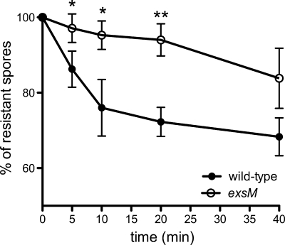
Bacillus cereus spores are assembled with a series of concentric layers that protect them from a wide range of environmental stresses. The outermost layer, or exosporium, is a bag-like structure that interacts with the environment and is composed of more than 20 proteins and glycoproteins. Here, we identified a new spore protein, ExsM, from a beta-mercaptoethanol extract of B. cereus ATCC 4342 spores. Subcellular localization of an ExsM-green fluorescent protein (GFP) protein revealed a dynamic pattern of fluorescence that follows the site of formation of the exosporium around the forespore. Under scanning electron microscopy, exsM null mutant spores were smaller and rounder than wild-type spores, which had an extended exosporium (spore length for the wt, 2.40 +/- 0.56 microm, versus that for the exsM mutant, 1.66 +/- 0.38 microm [P < 0.001]). Thin-section electron microscopy revealed that exsM mutant spores were encased by a double-layer exosporium, both layers of which were composed of a basal layer and a hair-like nap. Mutant exsM spores were more resistant to lysozyme treatment and germinated with higher efficiency than wild-type spores, and they had a delay in outgrowth. Insertional mutagenesis of exsM in Bacillus anthracis DeltaSterne resulted in a partial second exosporium and in smaller spores. In all, these findings suggest that ExsM plays a critical role in the formation of the exosporium.
Figures


Similar articles
-
The Exosporium Layer of Bacterial Spores: a Connection to the Environment and the Infected Host.Microbiol Mol Biol Rev. 2015 Dec;79(4):437-57. doi: 10.1128/MMBR.00050-15. Microbiol Mol Biol Rev. 2015. PMID: 26512126 Free PMC article. Review.
-
The Morphogenetic Protein CotE Positions Exosporium Proteins CotY and ExsY during Sporulation of Bacillus cereus.mSphere. 2021 Apr 21;6(2):e00007-21. doi: 10.1128/mSphere.00007-21. mSphere. 2021. PMID: 33883264 Free PMC article.
-
The BclB glycoprotein of Bacillus anthracis is involved in exosporium integrity.J Bacteriol. 2007 Sep;189(18):6704-13. doi: 10.1128/JB.00762-07. Epub 2007 Jul 20. J Bacteriol. 2007. PMID: 17644587 Free PMC article.
-
Coordinated Assembly of the Bacillus anthracis Coat and Exosporium during Bacterial Spore Outer Layer Formation.mBio. 2018 Nov 6;9(6):e01166-18. doi: 10.1128/mBio.01166-18. mBio. 2018. PMID: 30401771 Free PMC article.
-
The Bacillus anthracis Exosporium: What's the Big "Hairy" Deal?Microbiol Spectr. 2015 Oct;3(5). doi: 10.1128/microbiolspec.TBS-0021-2015. Microbiol Spectr. 2015. PMID: 26542035 Review.
Cited by
-
The Exosporium Layer of Bacterial Spores: a Connection to the Environment and the Infected Host.Microbiol Mol Biol Rev. 2015 Dec;79(4):437-57. doi: 10.1128/MMBR.00050-15. Microbiol Mol Biol Rev. 2015. PMID: 26512126 Free PMC article. Review.
-
Clostridium sordellii outer spore proteins maintain spore structural integrity and promote bacterial clearance from the gastrointestinal tract.PLoS Pathog. 2018 Apr 18;14(4):e1007004. doi: 10.1371/journal.ppat.1007004. eCollection 2018 Apr. PLoS Pathog. 2018. PMID: 29668758 Free PMC article.
-
Spore surface proteins of Brevibacillus laterosporus are involved in insect pathogenesis.Sci Rep. 2017 Mar 3;7:43805. doi: 10.1038/srep43805. Sci Rep. 2017. PMID: 28256631 Free PMC article.
-
Collagen-like glycoprotein BclS is involved in the formation of filamentous structures of the Lysinibacillus sphaericus exosporium.Appl Environ Microbiol. 2014 Nov;80(21):6656-63. doi: 10.1128/AEM.02238-14. Epub 2014 Aug 22. Appl Environ Microbiol. 2014. PMID: 25149519 Free PMC article.
-
The Regulation of Exosporium-Related Genes in Bacillus thuringiensis.Sci Rep. 2016 Jan 25;6:19005. doi: 10.1038/srep19005. Sci Rep. 2016. PMID: 26805020 Free PMC article.
References
-
- Ball, D. A., R. Taylor, S. J. Todd, C. Redmond, E. Couture-Tosi, P. Sylvestre, A. Moir, and P. A. Bullough. 2008. Structure of the exosporium and sublayers of spores of the Bacillus cereus family revealed by electron crystallography. Mol. Microbiol. 68:947-958. - PubMed
Publication types
MeSH terms
Substances
Grants and funding
LinkOut - more resources
Full Text Sources
Molecular Biology Databases
Research Materials
