

Mitochondrial dysfunction in the type 2 diabetic heart is associated with alterations in spatially distinct mitochondrial proteomes
- PMID: 20543078
- PMCID: PMC2930393
- DOI: 10.1152/ajpheart.00267.2010
Mitochondrial dysfunction in the type 2 diabetic heart is associated with alterations in spatially distinct mitochondrial proteomes
Abstract
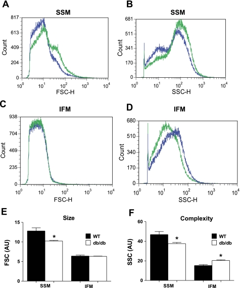
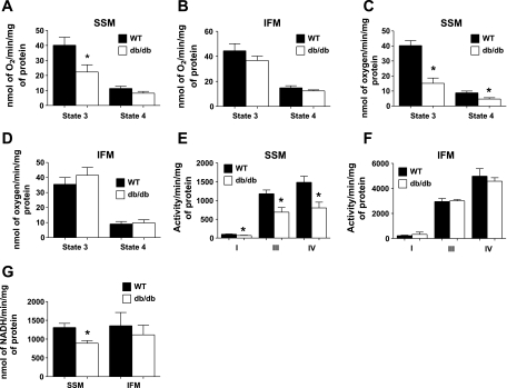
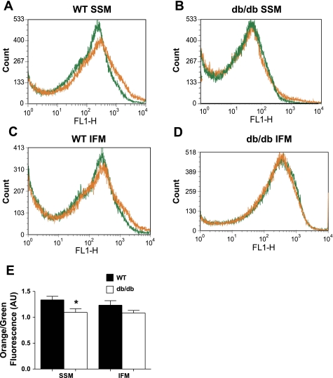
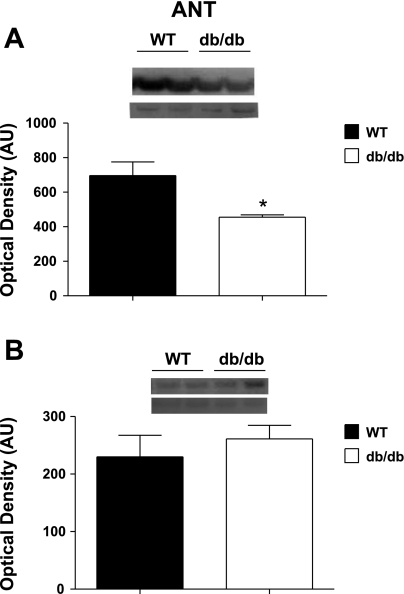
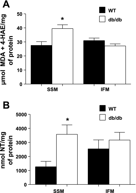
Cardiac complications and heart failure are the leading cause of death in type 2 diabetic patients. Mitochondrial dysfunction is central in the pathogenesis of the type 2 diabetic heart. However, it is unclear whether this dysfunction is specific for a particular subcellular region. The purpose of this study was to determine whether mitochondrial dysfunction in the type 2 diabetic heart is specific to a spatially distinct subset of mitochondria. We investigated mitochondrial morphology, function, and proteomic composition of subsarcolemmal mitochondria (SSM) and interfibrillar mitochondria (IFM) in 18-wk-old db/db mice. Oxidative damage was assessed in subpopulations through the measurement of lipid peroxidation byproducts and nitrotyrosine residues. Proteomic profiles and posttranslational modifications were assessed in mitochondrial subpopulations using iTRAQ and multi-dimensional protein identification technologies, respectively. SSM from db/db hearts had altered morphology, including a decrease in size and internal complexity, whereas db/db IFM were increased in internal complexity. Db/db SSM displayed decreased state 3 respiration rates, electron transport chain activities, ATP synthase activities, and mitochondrial membrane potential and increased oxidative damage, with no change in IFM. Proteomic assessment revealed a greater impact on db/db SSM compared with db/db IFM. Inner mitochondrial membrane proteins, including electron transport chain, ATP synthesis, and mitochondrial protein import machinery, were predominantly decreased. We provide evidence that mitochondrial dysfunction in the type 2 diabetic heart is associated with a specific subcellular locale. Furthermore, mitochondrial morphological and functional indexes are impacted differently during type 2 diabetic insult and may result from the modulation of spatially distinct mitochondrial proteomes.
Figures

Similar articles
-
Transgenic overexpression of mitofilin attenuates diabetes mellitus-associated cardiac and mitochondria dysfunction.J Mol Cell Cardiol. 2015 Feb;79:212-23. doi: 10.1016/j.yjmcc.2014.11.008. Epub 2014 Nov 22. J Mol Cell Cardiol. 2015. PMID: 25463274 Free PMC article.
-
Proteomic alterations of distinct mitochondrial subpopulations in the type 1 diabetic heart: contribution of protein import dysfunction.Am J Physiol Regul Integr Comp Physiol. 2011 Feb;300(2):R186-200. doi: 10.1152/ajpregu.00423.2010. Epub 2010 Nov 3. Am J Physiol Regul Integr Comp Physiol. 2011. PMID: 21048079 Free PMC article.
-
Diabetic cardiomyopathy-associated dysfunction in spatially distinct mitochondrial subpopulations.Am J Physiol Heart Circ Physiol. 2009 Feb;296(2):H359-69. doi: 10.1152/ajpheart.00467.2008. Epub 2008 Dec 5. Am J Physiol Heart Circ Physiol. 2009. PMID: 19060128 Free PMC article.
-
Physiological and structural differences in spatially distinct subpopulations of cardiac mitochondria: influence of cardiac pathologies.Am J Physiol Heart Circ Physiol. 2014 Jul 1;307(1):H1-14. doi: 10.1152/ajpheart.00747.2013. Am J Physiol Heart Circ Physiol. 2014. PMID: 24778166 Free PMC article. Review.
-
Mitochondrial membrane transporters and metabolic switch in heart failure.Heart Fail Rev. 2019 Mar;24(2):255-267. doi: 10.1007/s10741-018-9756-2. Heart Fail Rev. 2019. PMID: 30535838 Review.
Cited by
-
Transgenic overexpression of mitofilin attenuates diabetes mellitus-associated cardiac and mitochondria dysfunction.J Mol Cell Cardiol. 2015 Feb;79:212-23. doi: 10.1016/j.yjmcc.2014.11.008. Epub 2014 Nov 22. J Mol Cell Cardiol. 2015. PMID: 25463274 Free PMC article.
-
FIsetin Preserves Interfibrillar Mitochondria to Protect Against Myocardial Ischemia-Reperfusion Injury.Cell Biochem Biophys. 2022 Mar;80(1):123-137. doi: 10.1007/s12013-021-01026-4. Epub 2021 Aug 14. Cell Biochem Biophys. 2022. PMID: 34392494
-
Characterisation of the Myocardial Mitochondria Structural and Functional Phenotype in a Murine Model of Diabetic Cardiomyopathy.Front Physiol. 2021 Sep 1;12:672252. doi: 10.3389/fphys.2021.672252. eCollection 2021. Front Physiol. 2021. PMID: 34539423 Free PMC article.
-
Functional Implications of Cardiac Mitochondria Clustering.Adv Exp Med Biol. 2017;982:1-24. doi: 10.1007/978-3-319-55330-6_1. Adv Exp Med Biol. 2017. PMID: 28551779 Free PMC article. Review.
-
Role of the Mitochondrial Protein Import Machinery and Protein Processing in Heart Disease.Front Cardiovasc Med. 2021 Sep 28;8:749756. doi: 10.3389/fcvm.2021.749756. eCollection 2021. Front Cardiovasc Med. 2021. PMID: 34651031 Free PMC article. Review.
References
-
- Abdul-Ghani MA, Muller FL, Liu Y, Chavez AO, Balas B, Zuo P, Chang Z, Tripathy D, Jani R, Molina-Carrion M, Monroy A, Folli F, Van Remmen H, DeFronzo RA. Deleterious action of FA metabolites on ATP synthesis: possible link between lipotoxicity, mitochondrial dysfunction, and insulin resistance. Am J Physiol Endocrinol Metab 295: E678–E685, 2008 - PubMed
-
- Bornhovd C, Vogel F, Neupert W, Reichert AS. Mitochondrial membrane potential is dependent on the oligomeric state of F1F0-ATP synthase supracomplexes. J Biol Chem 281: 13990–13998, 2006 - PubMed
-
- Boudina S, Abel ED. Mitochondrial uncoupling: a key contributor to reduced cardiac efficiency in diabetes. Physiology (Bethesda) 21: 250–258, 2006 - PubMed
-
- Boudina S, Sena S, Theobald H, Sheng X, Wright JJ, Hu XX, Aziz S, Johnson JI, Bugger H, Zaha VG, Abel ED. Mitochondrial energetics in the heart in obesity-related diabetes: direct evidence for increased uncoupled respiration and activation of uncoupling proteins. Diabetes 56: 2457–2466, 2007 - PubMed
-
- Bradford MM. A rapid and sensitive method for the quantitation of microgram quantities of protein utilizing the principle of protein-dye binding. Anal Biochem 72: 248–254, 1976 - PubMed
Publication types
MeSH terms
Substances
Grants and funding
LinkOut - more resources
Full Text Sources
Medical
Molecular Biology Databases
Miscellaneous