. 2010 Jul 15;185(2):1205-14.
doi: 10.4049/jimmunol.1000039. Epub 2010 Jun 11.

Allergen-induced airway remodeling is impaired in galectin-3-deficient mice

Affiliations

Allergen-induced airway remodeling is impaired in galectin-3-deficient mice

Xiao Na Ge et al. J Immunol. .

Abstract

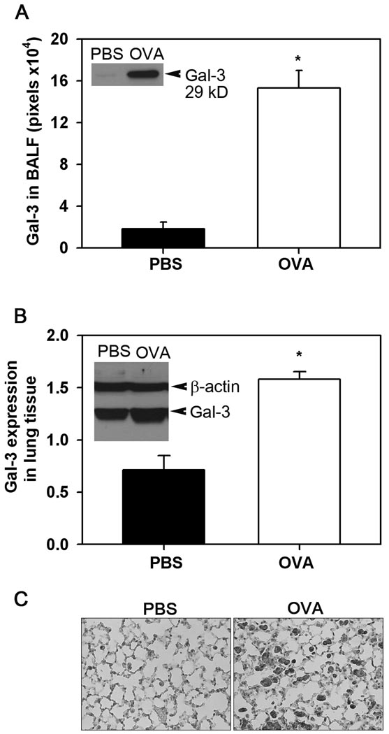
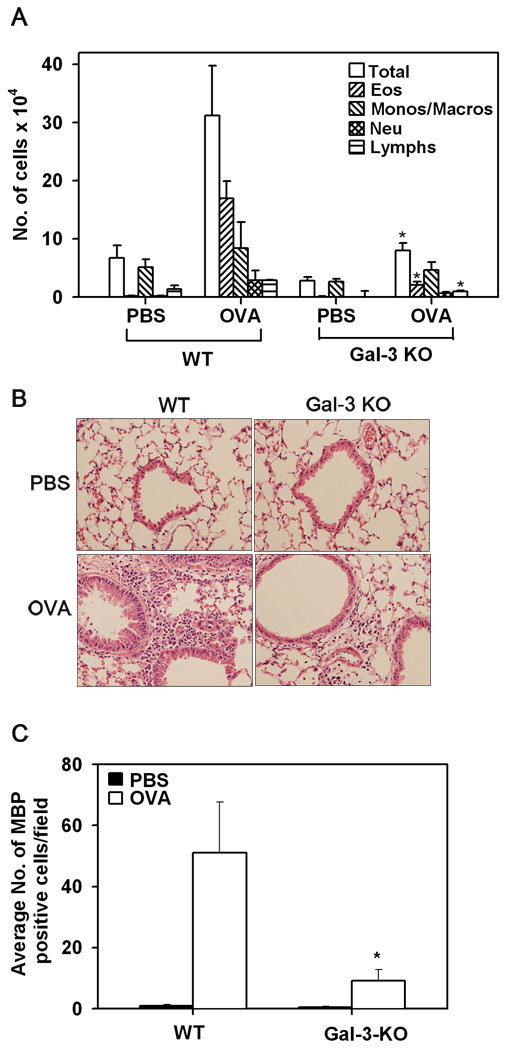
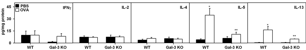
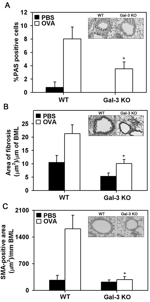
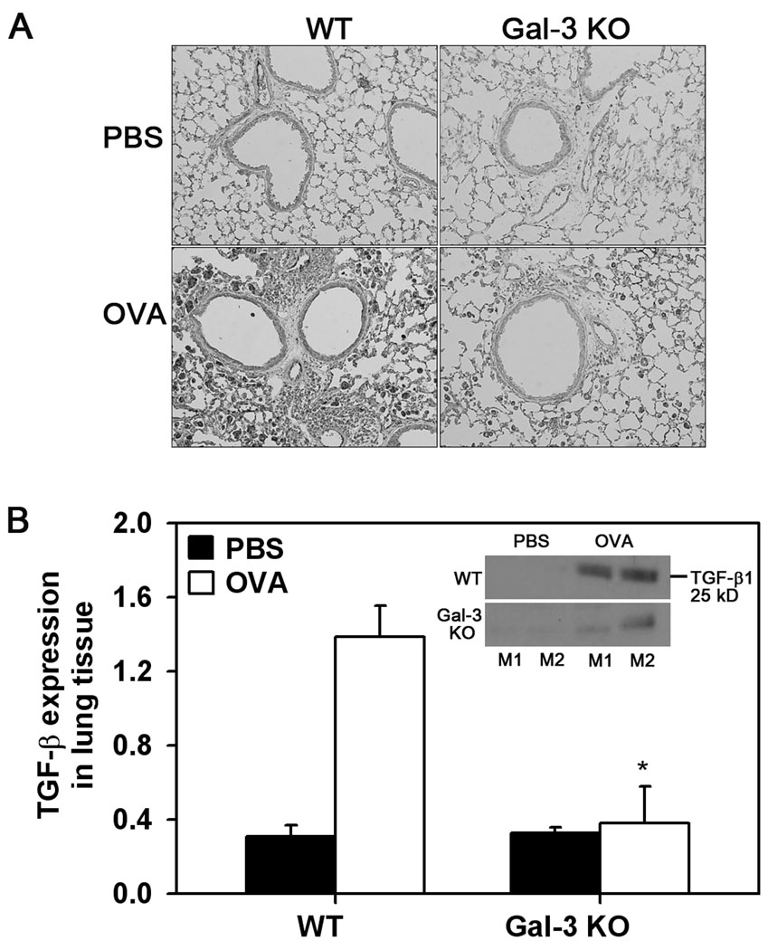
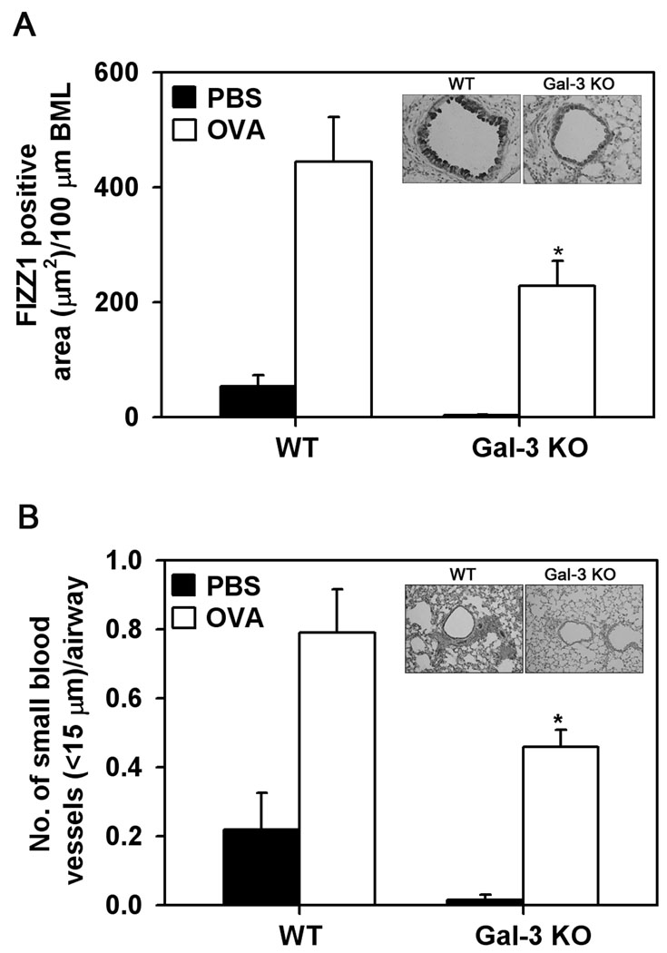
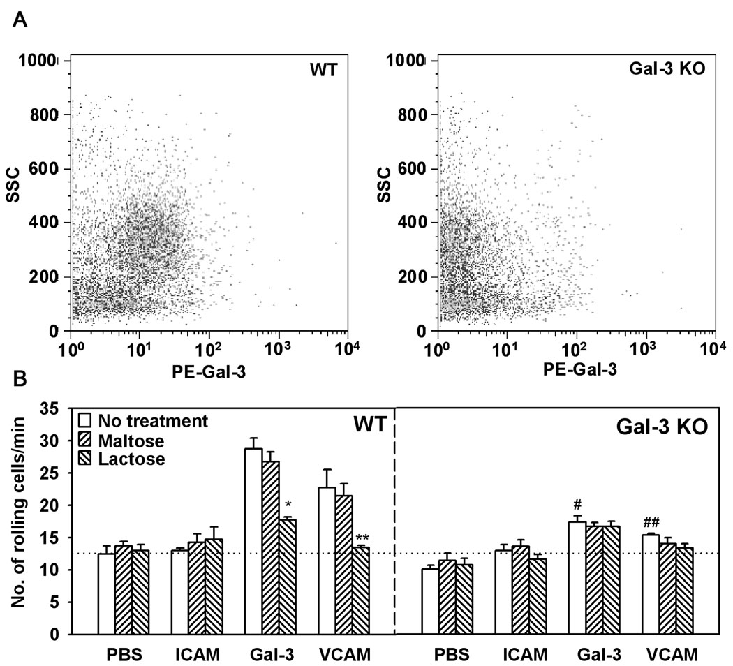
The role played by the beta-galactoside-binding lectin galectin-3 (Gal-3) in airway remodeling, a characteristic feature of asthma that leads to airway dysfunction and poor clinical outcome in humans, was investigated in a murine model of chronic allergic airway inflammation. Wild-type (WT) and Gal-3 knockout (KO) mice were subjected to repetitive allergen challenge with OVA up to 12 wk, and bronchoalveolar lavage fluid (BALF) and lung tissue collected after the last challenge were evaluated for cellular features associated with airway remodeling. Compared to WT mice, chronic OVA challenge in Gal-3 KO mice resulted in diminished remodeling of the airways with significantly reduced mucus secretion, subepithelial fibrosis, smooth muscle thickness, and peribronchial angiogenesis. The higher degree of airway remodeling in WT mice was associated with higher Gal-3 expression in the BALF as well as lung tissue. Cell counts in BALF and lung immunohistology demonstrated that eosinophil infiltration in OVA-challenged Gal-3 KO mice was significantly reduced compared with that WT mice. Evaluation of cellular mediators associated with eosinophil recruitment and airway remodeling revealed that levels of eotaxin-1, IL-5, IL-13, found in inflammatory zone 1, and TGF-beta were substantially lower in Gal-3 KO mice. Finally, leukocytes from Gal-3 KO mice demonstrated decreased trafficking (rolling) on vascular endothelial adhesion molecules compared with that of WT cells. Overall, these studies demonstrate that Gal-3 is an important lectin that promotes airway remodeling via airway recruitment of inflammatory cells, specifically eosinophils, and the development of a Th2 phenotype as well as increased expression of eosinophil-specific chemokines and profibrogenic and angiogenic mediators.


Figures

Fig. 1
Fig. 1. Gal-3 expression is up-regulated during chronic airway allergic inflammation
Gal-3 levels in BALF (A) and total lung tissue lysates (B) from control (PBS-exposed) and OVA-challenged WT mice were determined by Western blot analysis using rabbit antibodies against human Gal-3. Blots with lung tissue lysates were probed with HRP-conjugated anti-mouse β-actin to monitor levels of β-actin expression as an internal control. Bands were visualized on X-ray films, scanned and analyzed using ImageJ to quantitate the density (pixels) of the bands. Results (mean ± SE) were normalized for β-actin expression in the case of lung lysates (n = 4 mice/group). Inset in (A) and (B) show Gal-3 expression in BALF and lung lysate from representative control and OVA-challenged mice, respectively. *p < 0.05 compared with WT control mice. Immunohistochemical staining was performed on lung sections from control and OVA-challenged mice using rabbit antibodies against human Gal-3 as described in Materials and Methods. Representative images from each group at a magnification of ×200 are shown (C).
Fig. 2
Fig. 2. Cellular infiltration of the airways in response to chronic allergen challenge is inhibited in Gal-3 KO mice
BALF collected from control and OVA-challenged Gal-3 KO and WT mice 24 hours after the last challenge was evaluated for total as well as differential cell counts by microscopic evaluation of cytocentrifuged slides (n = 8 for control groups and 9 for OVA-challenged groups) and expressed as mean ± SE of the number of cells × 104 (A). Cellular infiltration of lung tissue was evaluated by H&E staining of paraformaldehyde-fixed lung tissue sections from control and allergen-challenged mice. Representative images from each group at a magnification of ×200 are shown (B). Lung tissue eosinophils were evaluated by immunohistochemical staining of lung sections with eosinophil-specific MBP using rat mAb against murine MBP. MBP-positive cells in five randomly selected non-overlapping microscopic fields were counted (magnification of ×400) and results were expressed as the average number (mean ± SE) of cells/field (n = 9 mice/group) (C). *p <0.05 when compared with WT OVA mice.
Fig. 3
Fig. 3. Eosinophils recruited to the airways in response to allergen challenge express Gal-3
BALF cells from OVA-challenged WT mice were dual stained with rat mAb against murine MBP and rabbit anti-human Gal-3 antibodies followed by FITC-conjugated goat anti-rat IgG and Rhodamine Red-X-conjugated goat anti-rabbit IgG to detect the bound primary antibodies. Cells were evaluated by confocal microscopy (magnification of ×600). Arrow heads indicate cells in a representative field that were positive for MBP as well as Gal-3 (A). BALF cells (non-permeabilized) from WT OVA-challenged mice were analyzed for surface Gal-3 expression by flow cytometry after treatment with Fc blocking antibody using total IgG purified from rabbit anti-Gal-3 serum as the primary antibody. Rabbit IgG was used as a control. PE-conjugated goat anti-rabbit IgG was used as the secondary antibody. Side-scatter profiles for control IgG (left) and anti-Gal-3 antibody (right) are shown (B).
Fig. 4
Fig. 4. Th2 cytokine levels are down-regulated in the lungs of Gal-3 KO mice in response to chronic allergen challenge
Key Th1 (IL-2 and IFN-γ) and Th2 (IL-4, IL-5 and IL-13) cytokine levels in lung lysates of control and chronic allergen-challenged WT and Gal-3 KO mice (n = 5–8/group) was determined by CBA. Results were expressed as mean ± SE of pg/mg protein for each cytokine. *p < 0.05 compared with WT PBS mice. **p <0.05 compared to WT OVA.
Fig. 5
Fig. 5. Allergen-challenged Gal-3 KO mice exhibit decreased eotaxin-1 levels
Eotaxin-1 levels in the BALF of control and OVA-challenged WT and Gal-3 KO mice (n= 7–9/group) were measured by ELISA. Concentration of eotaxin-1 in the BALF was determined against a generated standard curve (range 7.8 to 250 pg/ml) and expressed as pg/ml of BALF (mean ± SE) (A). Eotaxin-1 expression in lung tissue of control and chronic OVA-challenged WT and Gal-3 KO mice was evaluated by immunostaining using rat mAb against murine eotaxin-1. Representative images from each group at a magnification of ×100 are shown (B). Expression of eotaxin-1 mRNA level in lung tissue of control and OVA-challenged WT and Gal-3 KO mice (n = 7/group) was detected by qPCR. Results were expressed as mean ± SE of fold change (2−ΔΔCT) in eotaxin-1 expression after subtraction of internal β-actin control (C). *p < 0.05 compared with WT OVA mice.
Fig. 6
Fig. 6. Allergen-induced airway remodeling is attenuated in Gal-3 KO mice
Mucus secretion in the airways of control and chronic allergen-challenged WT and Gal-3 KO mice (n=6 mice/group) was quantitated by PAS staining of lung sections. The number of PAS-positive goblet cells was expressed as a percentage of the total number of epithelial cells in each airway (mean ± SE) (A). Peribroncial fibrosis was evaluated by staining lung sections with Masson’s trichrome stain, quantitated by image analysis using Image J and expressed as area of fibrosis (µm2)/µm basement membrane length (BML) (B). Thickness of the smooth muscle layer in lung sections was quantitated by immunohistochemical staining for SMA and expressed as SMA-positive area (µm2)/mm BML (C). The inset in each case shows representative images from OVA-challenged WT (left) and Gal-KO (right) mice at a magnification of ×200. *p < 0.05 when compared with WT OVA mice.
Fig. 7
Fig. 7. TGF-β1 expression is decreased in allergen-challenged Gal-3 KO mice
TGF-β1 expression in lung tissue of control and OVA-challenged WT mice and Gal-3 KO mice (n = 6/group) was detected by immunohistochemistry with polyclonal antibodies against TGF-β1. Representative images from each group at a magnification of ×100 are shown (A). Level of expression of mature TGF-β1 (25 kD) was quantitated from densitometric analysis of Western blots of lung tissue lysates from control and OVA-challenged WT and Gal-3 KO mice (n = 4/group) after normalizing for β-actin expression and expressed as mean ± SE (B). *p < 0.05 when compared with WT OVA mice.
Fig. 8
Fig. 8. Allergen-induced angiogenesis is inhibited in Gal-3 KO mice
FIZZ1 expression in lung tissue of control and OVA-challenged WT and Gal-3 KO mice (n = 6/group) was evaluated by immunohistochemistry using goat polyclonal antibodies against murine FIZZ1. Airway FIZZ1 expression was quantitated by image analysis using Image J and expressed as FIZZ1-positive area (µm2)/100 µm BML (A) Peribronchial angiogenesis was evaluated by immunohistochemical staining of lung sections with antibodies against CD31. The number of blood vessels with a diameter less than 15 µm in the area surrounding the airways (150 µm) was quantitated and expressed as the number of blood vessel per airway (B). Data represent mean ± SE. The inset shows representative images from OVA-challenged WT (left) and Gal-KO (right) mice at a magnification of ×200. *p < 0.05 when compared with WT OVA mice.
Fig. 9
Fig. 9. Bone marrow leukocytes from Gal-3 KO mice exhibit decreased rolling on endothelial adhesion molecules
Bone marrow leukocytes (non-permeabilized) from WT and Gal-3 KO mice were analyzed for surface Gal-3 expression by flow cytometry using total IgG purified from rabbit anti-Gal-3 serum (10 µg/ml) as the primary antibody after treatment with Fc blocking antibody. PE-conjugated goat anti-rabbit IgG (10 µg/ml) was used as the secondary antibody. Side-scatter profiles for bone marrow leukocytes from WT (left) and Gal-3 KO (right) mice are shown (A). Single cell suspensions of bone marrow leukocytes from WT and Gal-3 KO mice were infused into a flow chamber containing cover-slips coated with rmGal-3 or rmVCAM-1 at a flow rate of 1 ml/minute for 5 minutes. ICAM-1 was used as a negative control. In some experiments, bone marrow leukocytes were pre-incubated with lactose or maltose (as a control) at a concentration of 3 mM before infusion. Results were expressed as the number of rolling cells per minute (mean ± SE) (B). *p <0.01 versus rolling of untreated WT BM cells on rmGal-3; **p <0.01 versus rolling of untreated WT BM cells on rmVCAM-1; #p <0.01 versus rolling of untreated WT BM cells on rmGal-3; ## p <0.05 versus rolling of untreated WT BM cells on rmVCAM-1.

Similar articles

Cited by

References

    1. Bergeron C, Al-Ramli W, Hamid Q. Remodeling in asthma. Proc Am Thorac Soc. 2009;6:301–305. - PubMed
    1. Tagaya E, Tamaoki J. Mechanisms of airway remodeling in asthma. Allergol Int. 2007;56:331–340. - PubMed
    1. Mehrotra AK, Henderson WR., Jr The role of leukotrienes in airway remodeling. Curr Mol Med. 2009;9:383–391. - PubMed
    1. Doherty T, Broide D. Cytokines and growth factors in airway remodeling in asthma. Current Opinion in Immunology. 2007;19:676–680. - PubMed
    1. Sato S, Hughes RC. Regulation of secretion and surface expression of Mac-2, a galactoside-binding protein of macrophages. J Biol Chem. 1994;269:4424–4430. - PubMed

Publication types

MeSH terms