. 2010 Jul 15;185(2):1265-73.
doi: 10.4049/jimmunol.0902808. Epub 2010 Jun 11.

The relative timing of exposure to phagocytosable particulates and to osteoclastogenic cytokines is critically important in the determination of myeloid cell fate

Affiliations

The relative timing of exposure to phagocytosable particulates and to osteoclastogenic cytokines is critically important in the determination of myeloid cell fate

Douglas E James et al. J Immunol. .

Abstract

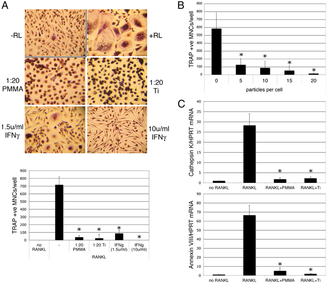
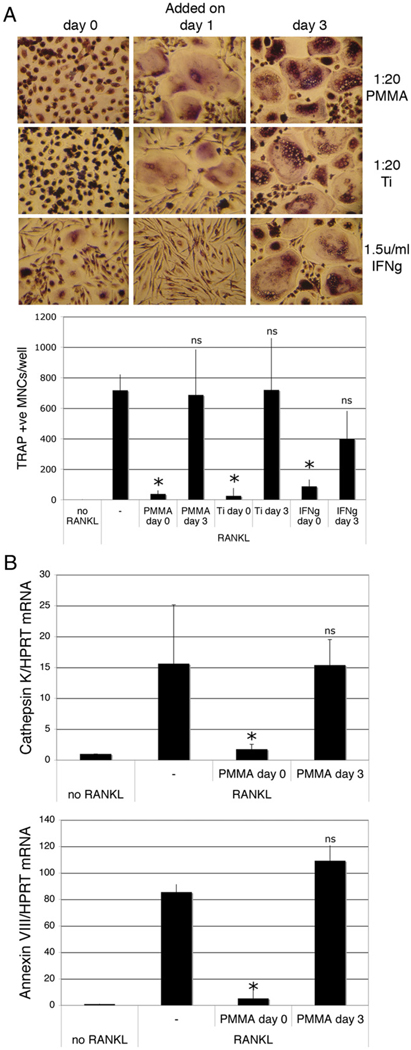
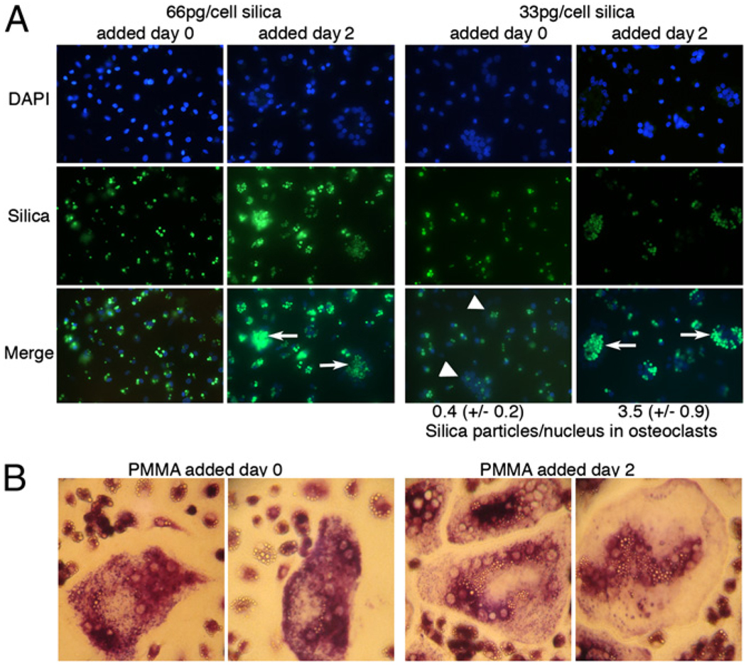
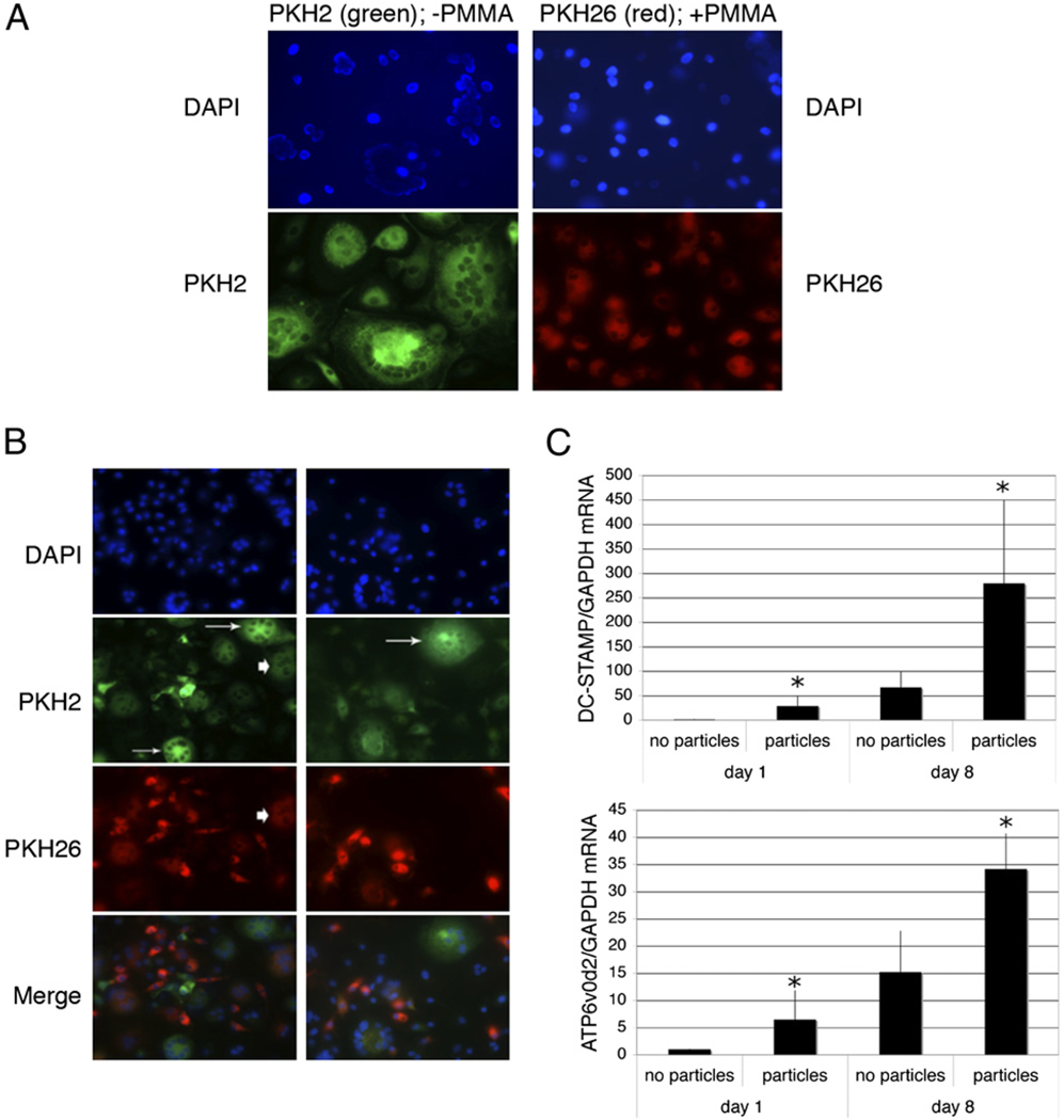
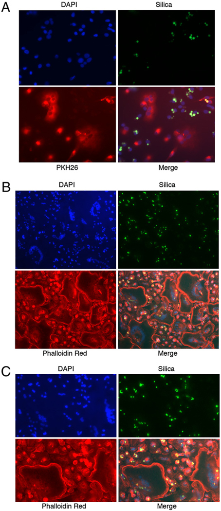
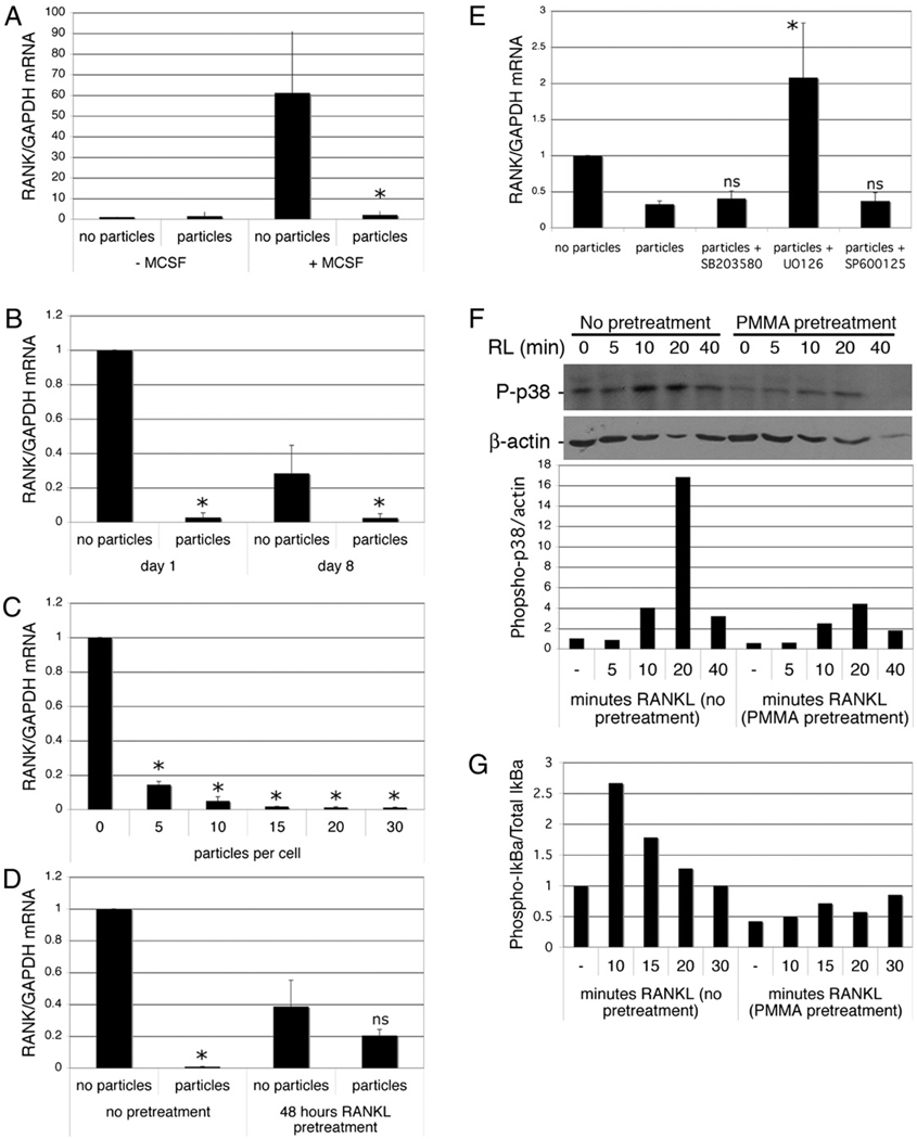
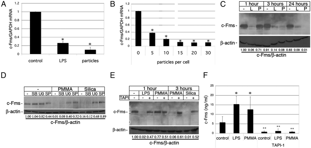
During granulomatous inflammatory reactions, myeloid cells can differentiate into activated phagocytic macrophages, wound-healing macrophages, foreign body giant cells, and bone-resorbing osteoclasts. Although it is appreciated that a variety of stimuli, including cytokines, cell-matrix interactions, and challenge with foreign materials can influence myeloid cell fate, little is known of how these signals integrate during this process. In this study, we have investigated the cross talk between receptor activator of NF-kappaB ligand (RANKL)-induced osteoclastogenesis and particle phagocytosis-induced activation of human monocytes. Understanding interconnected signals is of particular importance to disorders, such as periprosthetic osteolysis, in which granulomatous inflammation is initiated by particle phagocytosis in proximity to bone and leads to inflammatory bone loss. Using cell-based osteoclastogenesis and phagocytosis assays together with expression analysis of key regulators of osteoclastogenesis, we show in this study that phagocytosis of disease-relevant particles inhibits RANKL-mediated osteoclastogenesis of human monocytes. Mechanistically, phagocytosis mediates this effect by downregulation of RANK and c-Fms, the receptors for the essential osteoclastogenic cytokines RANKL and M-CSF. RANKL pretreatment of monocytes generates preosteoclasts that are resistant to RANK downregulation and committed to osteoclast formation, even though they retain phagocytic activity. Thus, the relative timing of exposure to phagocytosable particulates and to osteoclastogenic cytokines is critically important in the determination of myeloid cell fate.


Conflict of interest statement

Disclosures

The authors have no financial conflicts of interest.

Figures

FIGURE 1
FIGURE 1
Particle phagocytosis inhibits RANKL-mediated osteoclastogenesis. Preosteoclasts were cultured for 9 d in the presence of 25 ng/ml M-CSF in the presence or absence of RANKL (RL; 50 ng/ml). Particles of PMMA or titanium (Ti; 20 particles/cell) or IFN-γ (1.5 or 10 U/ml) were added at the same time as the RANKL. Following culture, cells were either stained for TRAP or processed for RNA extraction and qPCR. A, Representative images of TRAP-stained cells (original magnification ×50) and quantitation of TRAP-positive multinuclear (≥3 nuclei) cells. n = 3. B, Dose dependency of particle-mediated inhibition of RANKL-mediated osteoclastogenesis. n = 3. C, qPCR analysis of cathepsin K and Annexin VIII mRNA expression relative to GAPDH. n = 4. *p < 0.01 compared with RANKL alone.
FIGURE 2
FIGURE 2
RANKL pretreatment blunts particle phagocytosis-mediated inhibition of osteoclastogenesis. Preosteoclasts were cultured for 9 d in the presence of 25 ng/ml M-CSF and 50 ng/ml RANKL. Particles of PMMA or titanium (Ti; 20 particles/cell) or IFN-γ (1.5 U/ml) were added at the same time as the RANKL (day 0) or at various times after initial addition of RANKL. A, Representative TRAP staining of cells pretreated with RANKL for 0, 1, and 3 d and quantitation of TRAP-positive multinuclear cells (original magnification ×50). n = 3. B, qPCR for detection of cathepsin K and Annexin VIII mRNA of cells pretreated with RANKL for 0 and 3 d. n = 4. *p < 0.01 compared with RANKL alone.
FIGURE 3
FIGURE 3
RANKL-pretreated cells acquire both phagocytic and osteoclastogenic capacities. A, Preosteoclasts were cultured for 9 d in the presence of 25 ng/ml M-CSF and 50 ng/ml RANKL. Particles of green fluorescent silica were added at the same time as the RANKL (day 0) or 2 d after initial addition of RANKL. Cells were then analyzed by fluorescence microscopy (original magnification ×25). Arrows show osteoclasts formed in cells pretreated with RANKL; arrowheads show osteoclasts formed in cells without RANKL pretreatment. Particles per nucleus in osteoclasts were quantified as described in Materials and Methods. B, Preosteoclasts were cultured for 9 d in the presence of 25 ng/ml M-CSF and 50 ng/ml RANKL. Particles of PMMAwere added at the same time as the RANKL (day 0, 10 particles/cell) or 2 d after initial addition of RANKL (20 particles/cell). Osteoclastogenesis was determined by TRAP staining (original magnification ×100).
FIGURE 4
FIGURE 4
Particle phagocytosis prevents initiation of osteoclastogenesis but does not eliminate fusion with developing osteoclasts. A and B, Preosteoclasts were cytoplasmically labeled with PKH2 (green) or allowed to phagocytose PMMA particles and then labeled with PKH26 (red). The two cell populations were then cultured in the presence of 25 ng/ml M-CSF and 50 ng/ml RANKL either alone (A; original magnification ×50) or as cocultures (B; original magnification ×25). Equivalent results were obtained in three separate experiments. Osteoclasts formed exclusively from particle-free cells are indicated by arrows, and osteoclasts that have incorporated both particle-free and particle-containing cells are indicated with arrowheads. C, Preosteoclasts were cultured for 1 and 8 d with and without PMMA particles (20 particles/cell) in medium supplemented with 25 ng/ml M-CSF. RNA was extracted and DC-STAMP and ATP6v0d2 expression measured by qPCR. DC-STAMP and ATP6v0d2 mRNA levels expressed relative to GAPDH were normalized to the day 1 samples without particles. n = 5. *p < 0.05 compared with equivalent sample without particles.
FIGURE 5
FIGURE 5
Cells containing phagocytosed silica cannot initiate osteoclastogenesis, but may participate in cell fusion during later stages of osteoclast differentiation. Preosteoclasts were allowed to phagocytose green fluorescent silica particles and subsequently cocultured in the presence of 25 ng/ml M-CSF and 50 ng/ml RANKL with particle-free cells that had been labeled with PKH26 dye (A, red; original magnification ×50) or were unlabeled (B; original magnification ×25). Following culture, cells were labeled with phalloidin-Texas Red (B) and visualized by fluorescence microscopy. C shows a higher magnification (original magnification ×40) image of B. Equivalent results were obtained in two independent experiments.
FIGURE 6
FIGURE 6
Phagocytosis inhibits expression of RANK. A, Preosteoclasts were isolated and precultured overnight in the absence of M-CSF, then cultured with and without M-CSF (25 ng/ml) and PMMA (20 particles/cell). After 6 h, RNA was extracted and RANK expression measured by qPCR. RANK mRNA levels expressed relative to GAPDH were normalized to the cells treated without M-CSF or particles. n = 3. *p < 0.05 compared with sample with M-CSF and without PMMA. B, Preosteoclasts were cultured for 1 and 8 d with and without PMMA particles (20 particles/cell) in medium supplemented with 25 ng/ml M-CSF. RNA was extracted and RANK expression measured by qPCR. RANK mRNA levels expressed relative to GAPDH were normalized to the day 1 samples without particles. n = 5. *p < 0.01 compared with equivalent sample without particles. C, Dose dependency of particle-mediated repression of RANK expression in preosteoclasts 1 d after particle addition. n = 3. *p < 0.01 compared with equivalent sample without particles. D, Preosteoclasts were cultured for 1 d with and without PMMA particles (20 particles/cell) in medium supplemented with 25 ng/ml M-CSF and 50 ng/ml RANKL with or without a 48-h pretreatment with RANKL. n = 3. *p < 0.01 compared with equivalent sample without particles. E, Preosteoclasts were cultured for 6 h with and without PMMA particles (20 particles/cell) in medium supplemented with 25 ng/ml M-CSF. Where indicated, MAPK inhibitors were added 30 min prior to particles. RNA was extracted and RANK expression measured by qPCR. RANK mRNA levels expressed relative to GAPDH were normalized to the sample without particles. n = 3. *p < 0.05 compared with sample with particles but no inhibitors. F and G, Preosteoclasts were incubated with or without particles for 24 h, after which they were challenged with RANKL. Protein extracts were prepared and analyzed by immunoblotting for phosphorylated p38 and actin (F) or phosphorylated IκBα and total IκBα (G). Quantitation (as described in Materials and Methods) was normalized to control (cells without RANKL or particles). Results from one of two similar experiments are shown.
FIGURE 7
FIGURE 7
Phagocytosis inhibits expression of c-Fms. A, Preosteoclasts were cultured for 1 d with and without PMMA (20 particles/cell) or 100 ng/ml LPS in medium supplemented with 25 ng/ml M-CSF. RNA was extracted and c-Fms expression measured by qPCR. c-Fms mRNA levels expressed relative to GAPDH were normalized to the control sample without particles or LPS. n = 4. *p < 0.01 compared with control. B, Dose dependency of particle-mediated repression of c-Fms expression in preosteoclasts 1 d after particle addition. n = 3. *p < 0.01 compared with equivalent sample without particles. C, Preosteoclasts were treated with PMMA or LPS for 1, 3, or 24 h, and whole-cell protein lysates were analyzed by immunoblotting with anti–c-Fms Ab. Membranes were then reblotted with anti–β-actin Abs, and c-Fms levels were quantitated relative to β-actin. D, Preosteoclasts were preincubated with the MAPK inhibitors SB203580 (SB), U0216 (U0), and SP600125 (SP) for 30 min prior to addition of PMMA or silica (20 particles/cell). After 3 h, protein extracts were prepared and analyzed by immunoblotting as above. E, Preosteoclasts were pretreated with TAPI for 30 min prior to addition of LPS or particles. After 1 or 3 h, protein extracts were prepared and analyzed by immunoblotting. F, Preosteoclasts were pretreated with TAPI for 30 min and then treated with or without PMMA or LPS for 3 h, and c-Fms ectodomain shed into conditioned media was analyzed by ELISA. n = 3. *p < 0.01 compared with control; **p < 0.01 compared with equivalent samples without TAPI.

Similar articles

Cited by

References

    1. Boyle WJ, Simonet WS, Lacey DL. Osteoclast differentiation and activation. Nature. 2003;423:337–342. - PubMed
    1. Brodbeck WG, Anderson JM. Giant cell formation and function. Curr. Opin. Hematol. 2009;16:53–57. - PMC - PubMed
    1. Ross FP. M-CSF, c-Fms, and signaling in osteoclasts and their precursors. Ann. N. Y. Acad. Sci. 2006;1068:110–116. - PubMed
    1. Ha H, Lee JH, Kim HN, Kwak HB, Kim HM, Lee SE, Rhee JH, Kim HH, Lee ZH. Stimulation by TLR5 modulates osteoclast differentiation through STAT1/IFN-beta. J. Immunol. 2008;180:1382–1389. - PubMed
    1. Takami M, Kim N, Rho J, Choi Y. Stimulation by toll-like receptors inhibits osteoclast differentiation. J. Immunol. 2002;169:1516–1523. - PubMed

Publication types

MeSH terms