. 2010 Aug 20;285(34):25978-86.
doi: 10.1074/jbc.M110.142604. Epub 2010 Jun 11.

Discovery and Characterization of HemQ: an essential heme biosynthetic pathway component

Affiliations

Discovery and Characterization of HemQ: an essential heme biosynthetic pathway component

Tamara A Dailey et al. J Biol Chem. .

Abstract

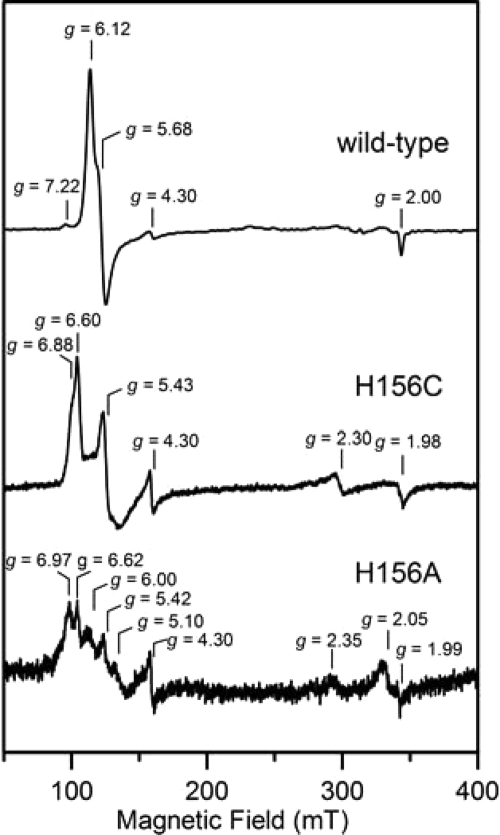
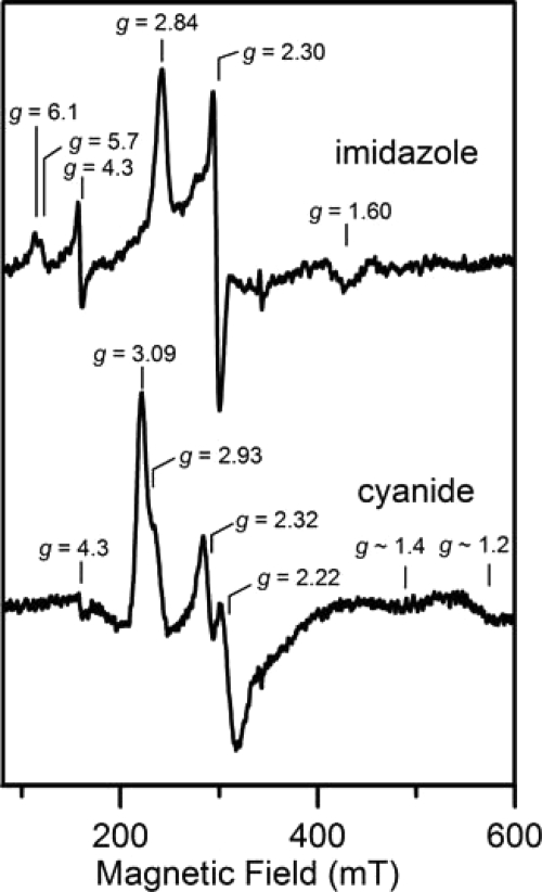
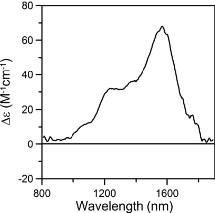
Here we identify a previously undescribed protein, HemQ, that is required for heme synthesis in Gram-positive bacteria. We have characterized HemQ from Bacillus subtilis and a number of Actinobacteria. HemQ is a multimeric heme-binding protein. Spectroscopic studies indicate that this heme is high spin ferric iron and is ligated by a conserved histidine with the sixth coordination site available for binding a small molecule. The presence of HemQ along with the terminal two pathway enzymes, protoporphyrinogen oxidase (HemY) and ferrochelatase, is required to synthesize heme in vivo and in vitro. Although the exact role played by HemQ remains to be characterized, to be fully functional in vitro it requires the presence of a bound heme. HemQ possesses minimal peroxidase activity, but as a catalase it has a turnover of over 10(4) min(-1). We propose that this activity may be required to eliminate hydrogen peroxide that is generated by each turnover of HemY. Given the essential nature of heme synthesis and the restricted distribution of HemQ, this protein is a potential antimicrobial target for pathogens such as Mycobacterium tuberculosis.


Figures

FIGURE 1.
FIGURE 1.
Genomic organization of the heme biosynthetic pathway genes in P. acnes (adapted from NCBI Entrez Gene). The entire heme biosynthetic pathway in this organism is organized into two uber operons (58 and 59). The numbers above the line denote the sequence position of this operon. The letters signify the hem gene names and correspond to the following: hemA, glutamyl tRNA reductase; hemB, porphobilinogen synthase (5-aminolevulinate dehydratase); hemC, hydroxybilane synthase (porphobilinogen deaminase); hemD, uroporphyrinogen synthase; hemE, uroporphyrinogen decarboxylase; hemL, glutamate 1-semialdehyde aminotransferase; hemY, protoporphyrinogen oxidase; and hemH/Q, ferrochelatase-hemQ fusion.
FIGURE 2.
FIGURE 2.
ClustalW analysis of select COG3253 proteins. The segment shown spans from residues number 71 to 186 of the B. subtilis protein. The sequences are boxed to show HemQ sequences from Gram-positive bacteria on top, HemQ sequences of Actinobacteria in the middle, and chlorite dismutases on the bottom.
FIGURE 3.
FIGURE 3.
Sephacryl S-300 size exclusion chromatography and SDS-PAGE of purified HemQ from M. tuberculosis. Details are under “Experimental Procedures.” The 1st eluted peak corresponds to the column void volume. The major protein peak elutes at a position consistent with a soluble globular protein of ∼200,000 daltons. The inset shows purified HemQ adjacent to molecular weight markers (Bio-Rad Precision Plus, where the darker bands correspond to 75,000, 50,000, and 25,000, respectively) in an SDS-polyacrylamide gel. HemQ migrates with an apparent molecular weight of 26,000.
FIGURE 4.
FIGURE 4.
UV-visible absorption spectra of M. tuberculosis HemQ. The figure shows the spectra of HemQ as purified in the presence of 250 mm imidazole with (solid line) and without (dashed line) added hemin. The base line of the two scans is shifted for clarity.
FIGURE 5.
FIGURE 5.
Titration of apo-HemQ with hemin. Purified HemQ (19.5 μm) was repetitively scanned following successive additions of freshly prepared hemin (100 μm stock solution in 1% (w/v) Triton X-100). Absorbance at 410 nm was plotted against the final concentration of hemin added.
FIGURE 6.
FIGURE 6.
Stimulation of HemY activity by HemQ. HemY from P. acnes was assayed as described in the text with HemQ that did not have added heme (lower curve) and with heme-loaded HemQ (upper curve).
FIGURE 7.
FIGURE 7.
Heme synthesis from protoporphyrinogen IX and ferrous iron. The bar graph shows heme formation in vitro by 1 nmol of HemY and HemH. When heme-loaded HemQ was employed in the assay, the amount of heme (present in holo-HemQ) added was quantitated and accounted for less than 5% of total heme in assays with heme forming activity. In one experiment (far right) HemQ was replaced with catalase, and in one experiment protoporphyrin, rather than the porphyrinogen, was supplied. In neither of these instances was heme formation observed.
FIGURE 8.
FIGURE 8.
UV-visible absorption spectra of heme-reconstituted wild-type (solid line), H156C (dot-dashed line), and H156A (dashed line) M. tuberculosis HemQ after dialysis to remove imidazole. The spectra have been normalized to give the same absorbance at 280 nm.
FIGURE 9.
FIGURE 9.
EPR spectra of heme-reconstituted wild-type, H156C, and H156A M. tuberculosis HemQ after dialysis to remove imidazole. The EPR spectra were recorded at 10 K with 1 milliwatt microwave power using a microwave frequency of 9.60 GHz and a modulation amplitude of 0.65 mT.
FIGURE 10.
FIGURE 10.
EPR spectra of heme-reconstituted M. tuberculosis HemQ in the presence of 250-fold excess of imidazole and a 25-fold excess of sodium cyanide. The EPR spectra were recorded at 10 K with 5 milliwatt microwave power using a microwave frequency of 9.60 GHz and a modulation amplitude of 0.65 mT.
FIGURE 11.
FIGURE 11.
Near-IR low temperature MCD spectrum of heme-reconstituted M. tuberculosis HemQ in the presence of 250-fold excess of imidazole. The spectrum was recorded with an applied magnetic field of 6 T at 1.8 K.

Similar articles

Cited by

References

    1. Ponka P. (1999) Am. J. Med. Sci. 318, 241–256 - PubMed
    1. Ortiz de Orué Lucana D., Groves M. R. (2009) Amino Acids 37, 479–486 - PubMed
    1. Roberts G. P., Kerby R. L., Youn H., Conrad M. (2005) J. Inorg. Biochem. 99, 280–292 - PubMed
    1. Spiro T. (2008) ACS Chem. Biol. 3, 673–675 - PMC - PubMed
    1. Schläfli P., Borter E., Spielmann P., Wenger R. H. (2009) Cell. Mol. Life Sci. 66, 876–883 - PMC - PubMed

Publication types

MeSH terms

LinkOut - more resources