

Single-spanning transmembrane domains in cell growth and cell-cell interactions: More than meets the eye?
- PMID: 20543559
- PMCID: PMC2900628
- DOI: 10.4161/cam.4.2.12430
Single-spanning transmembrane domains in cell growth and cell-cell interactions: More than meets the eye?
Abstract
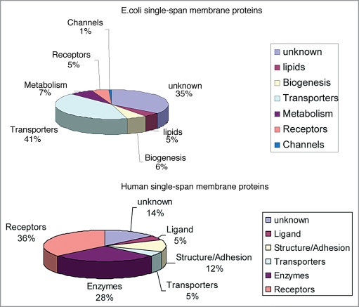
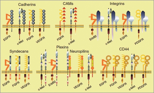
As a whole, integral membrane proteins represent about one third of sequenced genomes, and more than 50% of currently available drugs target membrane proteins, often cell surface receptors. Some membrane protein classes, with a defined number of transmembrane (TM) helices, are receiving much attention because of their great functional and pharmacological importance, such as G protein-coupled receptors possessing 7 TM segments. Although they represent roughly half of all membrane proteins, bitopic proteins (with only 1 TM helix) have so far been less well characterized. Though they include many essential families of receptors, such as adhesion molecules and receptor tyrosine kinases, many of which are excellent targets for biopharmaceuticals (peptides, antibodies, et al.). A growing body of evidence suggests a major role for interactions between TM domains of these receptors in signaling, through homo and heteromeric associations, conformational changes, assembly of signaling platforms, etc. Significantly, mutations within single domains are frequent in human disease, such as cancer or developmental disorders. This review attempts to give an overview of current knowledge about these interactions, from structural data to therapeutic perspectives, focusing on bitopic proteins involved in cell signaling.
Figures
Similar articles
-
Evidence for new homotypic and heterotypic interactions between transmembrane helices of proteins involved in receptor tyrosine kinase and neuropilin signaling.J Mol Biol. 2014 Dec 12;426(24):4099-4111. doi: 10.1016/j.jmb.2014.10.007. Epub 2014 Oct 12. J Mol Biol. 2014. PMID: 25315821
-
Intramembrane helix-helix association in oligomerization and transmembrane signaling.Annu Rev Biophys Biomol Struct. 1992;21:223-42. doi: 10.1146/annurev.bb.21.060192.001255. Annu Rev Biophys Biomol Struct. 1992. PMID: 1326354 Review.
-
Structure elucidation of dimeric transmembrane domains of bitopic proteins.Cell Adh Migr. 2010 Apr-Jun;4(2):284-98. doi: 10.4161/cam.4.2.11930. Epub 2010 May 1. Cell Adh Migr. 2010. PMID: 20421711 Free PMC article. Review.
-
Transmembrane helix-helix interactions involved in ErbB receptor signaling.Cell Adh Migr. 2010 Apr-Jun;4(2):299-312. doi: 10.4161/cam.4.2.11191. Epub 2010 Apr 13. Cell Adh Migr. 2010. PMID: 20212358 Free PMC article. Review.
-
Dynamic helix interactions in transmembrane signaling.Cell. 2006 Nov 3;127(3):447-50. doi: 10.1016/j.cell.2006.10.016. Cell. 2006. PMID: 17081964
Cited by
-
Cryo-electron Microscopic Analysis of Single-Pass Transmembrane Receptors.Chem Rev. 2022 Sep 14;122(17):13952-13988. doi: 10.1021/acs.chemrev.1c01035. Epub 2022 Jun 17. Chem Rev. 2022. PMID: 35715229 Free PMC article. Review.
-
Lateral organization of biological membranes: role of long-range interactions.Eur Biophys J. 2013 Dec;42(11-12):843-50. doi: 10.1007/s00249-013-0933-x. Epub 2013 Oct 26. Eur Biophys J. 2013. PMID: 24158717
-
Transmembrane Interactions of Full-length Mammalian Bitopic Cytochrome-P450-Cytochrome-b5 Complex in Lipid Bilayers Revealed by Sensitivity-Enhanced Dynamic Nuclear Polarization Solid-state NMR Spectroscopy.Sci Rep. 2017 Jun 23;7(1):4116. doi: 10.1038/s41598-017-04219-1. Sci Rep. 2017. PMID: 28646173 Free PMC article.
-
Inhibition of PlexA1-mediated brain tumor growth and tumor-associated angiogenesis using a transmembrane domain targeting peptide.Oncotarget. 2016 Sep 6;7(36):57851-57865. doi: 10.18632/oncotarget.11072. Oncotarget. 2016. PMID: 27506939 Free PMC article.
-
Membranome: a database for proteome-wide analysis of single-pass membrane proteins.Nucleic Acids Res. 2017 Jan 4;45(D1):D250-D255. doi: 10.1093/nar/gkw712. Epub 2016 Aug 10. Nucleic Acids Res. 2017. PMID: 27510400 Free PMC article.
References
-
- Stevens TJ, Arkin IT. Do more complex organisms have a greater proportion of membrane proteins in their genomes? Proteins. 2000;39:417–420. - PubMed
-
- Fagerberg L, Jonasson K, von Heijne G, Uhlen M, Berglund L. Prediction of the human membrane proteome. Proteomics. 2010;10:1–9. - PubMed
-
- Glukhov E, Burrows LL, Deber CM. Membrane interactions of designed cationic antimicrobial peptides: The two thresholds. Biopolymers. 2008;89:360–371. - PubMed
Publication types
MeSH terms
Substances
LinkOut - more resources
Full Text Sources
Other Literature Sources
Research Materials