. 2010 Jul 1;9(13):2640-6.
doi: 10.4161/cc.9.13.12120.

The SUMO pathway functions in mouse oocyte maturation

Affiliations

The SUMO pathway functions in mouse oocyte maturation

Zhen-Bo Wang et al. Cell Cycle. .

Abstract

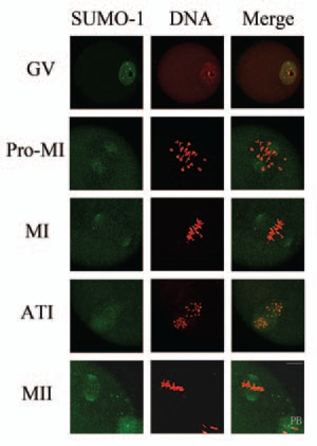
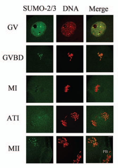
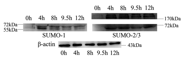
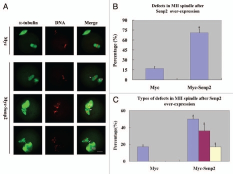
Sumoylation is an important post-translational modification in which SUMO (small ubiquitin-related modifier) proteins are bonded covalently to their substrates. Studies on the roles of sumoylation in cell cycle regulation have been emerging in both mitosis from yeast to mammals and meiosis in budding yeast, but the functions of sumoylation in mammalian meiosis, especially in oocyte meiotic maturation are not well known. Here, we examined the localization and expression of SUMO-1 and SUMO-2/3, the two basic proteins in the sumoylation pathway and investigated their roles through over-expression of Senp2 during mouse oocyte maturation. Immunofluorescent staining revealed differential patterns of SUMO-1 and SUMO-2/3 localization: SUMO-1 was localized to the spindle poles in prometaphase I, MI and MII stages, around the separating homologues in anaphase I and telophase I stages of first meiosis, while SUMO-2/3 was mainly concentrated near centromeres during mouse oocyte maturation. Immunoblot analysis uncovered the different expression profiles of SUMO-1 and SUMO-2/3 modified proteins during mouse oocyte maturation. Over-expression of Senp2, a SUMO-specific isopeptidase, caused changes of SUMO-modified proteins and led to defects in MII spindle organization in mature eggs. These results suggest that the SUMO pathway may play an indispensable role during mouse oocyte meiotic maturation.


Figures

Figure 1
Figure 1
Subcellular localization of SUMO-1 during mouse oocyte maturation. Oocytes were fixed at 0 h, 4 h, 8 h, 9.5 h and 12 h of culture and then stained with a mouse monoclonal antibody specific for SUMO-1 and counterstained with the fluorescent dye propidium Iodide (PI) to visualize DNA. Green, SUMO-1; red, DNA; GV, oocytes at germinal vesicle stage; Pro-MI, oocytes at first prometaphase; MI, oocytes at first metaphase; ATI, oocytes at first anaphase; MII, oocytes at second metaphase. PB, first polar body; Bar = 20 µm. Experiments were repeated at least three times and representative images are shown.
Figure 2
Figure 2
Subcellular localization of SUMO-2/3 during mouse oocyte maturation. Oocytes at different stages (0 h, 2 h, 8 h, 9.5 h and 12 h of culture, respectively) were immunolabeled with a mouse monoclonal antibody specific to SUMO-2/3 (green). GV, oocytes at germinal vesicle stage; GVBD, oocytes after germinal vesicle breakdown; MI, oocytes at first metaphase; ATI, oocytes at first anaphase; MII, oocytes at second metaphase. Each sample was counterstained with PI (red) to visualize DNA. PB, first polar body; Bar = 20 µm. Experiments were repeated at least three times and representative images are shown.
Figure 3
Figure 3
Expression of SUMO-1 and SUMO-2/3 modified proteins during mouse oocyte maturation. Samples were collected after oocyte culture for 0, 4, 8, 9.5 and 12 h, corresponding to the GV, prometaphase I, metaphase I, anaphase I, metaphase II stages, respectively. Proteins from a total of 250 oocytes were loaded for each sample. Experiments were repeated three times.
Figure 4
Figure 4
Overexpression of Senp2 resulted in changes of SUMO modified proteins. (A) Myc-Senp2 protein was expressed in mouse oocytes. Myc-Senp2 mRNA microinjected GV oocytes were incubated in M16 medium containing 2.5 µM milrinone for 5 h and collected immediately; the lysates were then analyzed by immunoblot with mouse monoclonal anti-myc antibody. Equal numbers of oocytes microinjected with Myc mRNA was analyzed as a control. (B) Germinal vesicle oocytes were microinjected with the control Myc mRNA and Myc-Senp2 mRNA, respectively. After microinjection, oocytes were incubated in M16 medium with 2.5 µM milrinone for 5 h, then transferred to milrinone-free M16 medium for 10 h and collected, followed by immunoblot analysis with SUMO-1 or SUMO-2/3 specific mouse monoclonal antibodies. Three independent experiments were performed.
Figure 5
Figure 5
Overexpression of Senp2 impaired spindle organization in MII mature eggs. (A) MII spindle morphologies in Myc mRNA and Myc-Senp2 mRNA microinjection groups. After microinjection, GV oocytes were incubated in M16 medium with 2.5 µM milrinone for 5 h, then transferred to milrinone-free M16 medium for 14 h to fully mature into MII eggs and fixed immediately, followed by immunoflurescent analysis with anti-α-tubulin-FITC antibody (green) and PI (red). In the Myc mRNA microinjection group, normal MII spindles formed. In the Myc-Senp2 mRNA microinjection group, several forms of morphologically aberrant MII spindles were seen in mature eggs. Arrow head, multipolar spindle; arrows, microtubule bundles connecting MII spindles and the first polar bodies. PB, first polar body, Bar = 20 µm. Three independent experiments were performed and representative images are shown. (B) The rate of abnormal MII spindles was recorded in the Myc mRNA microinjection group and the Myc-Senp2 mRNA microinjection group. Data are presented as mean percentage (mean ± SEM) from three independent experiments *p < 0.05. (C) The rates of the main forms of defects in MII spindles were recorded in the Myc mRNA microinjection group and the Myc-Senp2 mRNA microinjection group. Blue: MII spindles with defective morphology; purple: MII spindles with branched microtubule bundles connecting the first polar body; Yellow: MII spindles with both of the above defects. Data are presented as mean percentage (mean ± SEM) from three independent experiments *p < 0.05.

References

    1. Di Bacco A, Ouyang J, Lee HY, Catic A, Ploegh H, Gill G. The SUMO-specific protease SENP5 is required for cell division. Mol Cell Biol. 2006;26:4489–4498. - PMC - PubMed
    1. Geiss-Friedlander R, Melchior F. Concepts in sumoylation: a decade on. Nat Rev Mol Cell Biol. 2007;8:947–956. - PubMed
    1. Zhao J. Sumoylation regulates diverse biological processes. Cell Mol Life Sci. 2007;64:3017–3033. - PMC - PubMed
    1. Meulmeester E, Melchior F. Cell biology: SUMO. Nature. 2008;452:709–711. - PubMed
    1. Mukhopadhyay D, Dasso M. Modification in reverse: the SUMO proteases. Trends Biochem Sci. 2007;32:286–295. - PubMed

Publication types

Substances

LinkOut - more resources