. 2010 Jul 21;29(14):2368-80.
doi: 10.1038/emboj.2010.124. Epub 2010 Jun 11.

The C-terminal alpha-alpha superhelix of Pat is required for mRNA decapping in metazoa

Affiliations

The C-terminal alpha-alpha superhelix of Pat is required for mRNA decapping in metazoa

Joerg E Braun et al. EMBO J. .

Abstract

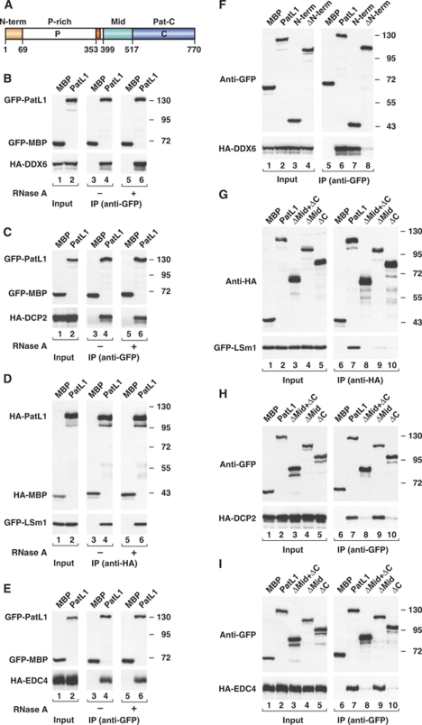
Pat proteins regulate the transition of mRNAs from a state that is translationally active to one that is repressed, committing targeted mRNAs to degradation. Pat proteins contain a conserved N-terminal sequence, a proline-rich region, a Mid domain and a C-terminal domain (Pat-C). We show that Pat-C is essential for the interaction with mRNA decapping factors (i.e. DCP2, EDC4 and LSm1-7), whereas the P-rich region and Mid domain have distinct functions in modulating these interactions. DCP2 and EDC4 binding is enhanced by the P-rich region and does not require LSm1-7. LSm1-7 binding is assisted by the Mid domain and is reduced by the P-rich region. Structural analysis revealed that Pat-C folds into an alpha-alpha superhelix, exposing conserved and basic residues on one side of the domain. This conserved and basic surface is required for RNA, DCP2, EDC4 and LSm1-7 binding. The multiplicity of interactions mediated by Pat-C suggests that certain of these interactions are mutually exclusive and, therefore, that Pat proteins switch decapping partners allowing transitions between sequential steps in the mRNA decapping pathway.


Conflict of interest statement

The authors declare that they have no conflict of interest.

Figures

Figure 1
Figure 1
PatL1 coimmunoprecipitates DDX6/RCK, LSm1, DCP2 and EDC4. (A) Pat proteins contain a conserved N-term sequence, a P-rich region, a Mid domain and Pat-C. Amino-acid positions at fragment boundaries are indicated for human PatL1. Red box: conserved sequence motif in the P-rich region. (BI) GFP- and HA-tagged proteins were coexpressed in human cells as indicated. Cell lysates were immunoprecipitated using anti-GFP or anti-HA antibodies. GFP- or HA-tagged maltose binding protein (MBP) served as a negative control. In lanes 5–6 of (BE), cell lysates were treated with RNase A before immunoprecipitation. Inputs and immunoprecipitates were analysed by western blotting using anti-GFP and anti-HA antibodies.
Figure 2
Figure 2
Pat-C is required for binding to DCP2, EDC4 and LSm1–7. (AC) GFP- and HA-tagged proteins were coexpressed in human cells as indicated. Cell lysates were immunoprecipitated using anti-GFP or anti-HA antibodies and analysed as described in Figure 1. Numbers in italics below the lanes in (A, B) represent coimmunoprecipitation efficiencies relative to wild-type PatL1. Values take into account differences in protein expression levels in the inputs and the relative amount of PatL1 proteins in the immunoprecipitates. (D) HA-tagged wild-type HPat and fragments were expressed in D. melanogaster S2 cells together with GFP-LSm1. Cell lysates were immunoprecipitated using anti-HA antibodies. Asterisks indicate cross-reactivity of the primary antibodies with an endogenous protein (input panels).
Figure 3
Figure 3
Pat-C is required for PatL1 incorporation into active decapping complexes. (AD) GFP-tagged proteins were expressed in human cells. Cell lysates were immunoprecipitated using anti-GFP antibodies. The immunoprecipitates were tested for decapping activity using in vitro synthesized 32P-labelled capped mRNA. Samples corresponding to (A, C) were analysed by western blotting in (B, D), respectively, to ensure that equivalent amounts of proteins were present in the decapping assay.
Figure 4
Figure 4
Pat-C is required for PatL1 accumulation in P-bodies. (AH) Representative confocal fluorescent micrographs of fixed human HeLa cells expressing wild-type GFP-PatL1 or the mutants indicated on the left. Cells were stained with antibodies cross-reacting with EDC4 and a nuclear human antigen (Kedersha and Anderson, 2007). The merged images show the GFP signal in green and the EDC4 signal in red. The fraction of cells exhibiting a staining identical to that shown in the representative panel was determined by scoring at least 100 cells in each of the three independent transfections performed per protein. Scale bar: 10 μm.
Figure 5
Figure 5
Structure of human Pat-C (Form II, chain A). (A) Ribbon diagram in two orientations related by a 180° rotation along a vertical axis. Loop L(α8–α9) is replaced by a Gly-Ser linker in Pat-C-Δ-loop construct. Structural representations were generated using Pymol (http://www.pymol.org). (B) Surface representation, coloured by sequence conservation, comparing six species (Supplementary Figure S2B). Colour ramp by identity: orange (100%) to white (0%). Highly conserved solvent exposed residues are labelled. (C) Electrostatic potentials are mapped onto the molecular surface of Pat-C and contoured from −10 kT/e (red) to 10 kT/e (blue). Residues contributing to the positively charged surface patch are labelled. (D) Ribbon representation of six Pat-C molecules resulting from the three independent crystal forms. Green (Form I, chain A), dark blue (Form II, chain A), light blue (Form II, chain B), orange (Form III, chain A), red (Form III, chain B), yellow (Form III, chain C). (E) Close-up view (40° rotated along a horizontal axis) on the helical protrusion of the six structurally most divergent Pat-C molecules. Colours are as in (D). (F) Close-up on Form I, chain A, showing two water molecules buried in the helical protrusion. Residues adjacent to the buried water molecules are labelled and shown as sticks with carbons in green, nitrogens in blue and oxygens in red. Hydrogen bonds are shown as dashed lines.
Figure 6
Figure 6
A basic and conserved surface on Pat-C enables PatL1 to interact with DCP2, EDC4 and the LSm1–7 ring. (AD) GFP- and HA-tagged proteins were coexpressed in human cells as indicated. Cell lysates were immunoprecipitated using anti-GFP or anti-HA antibodies and analysed as described in Figure 1.
Figure 7
Figure 7
Pat-C is required for decapping in vivo. (A) Pat-C conserved residues are required for HPat to interact with the LSm1–7 complex. GFP- and HA-tagged proteins were coexpressed in D. melanogaster S2 cells as indicated. Cell lysates were immunoprecipitated using a monoclonal anti-HA antibody. HA-GST (Glutathion-S-Transferase) served as negative control. Inputs and immunoprecipitates were analysed as described in Figure 1. (BD) Control S2 cells (treated with GFP dsRNA) or cells codepleted of HPat and Me31B were cotransfected with a mixture of three plasmids: one expressing the F-Luc-5BoxB reporter, another expressing λN-HA-GW182 or the λN-HA peptide and a third expressing Renilla luciferase (R-Luc). Plasmids (5 ng) expressing HA-MBP, wild-type HA-HPat, HPatΔC or Mut2 were included in the transfection mixtures, as indicated. RNA samples were analysed by northern blot. F-Luc-5BoxB mRNA levels were normalized to those of the Renilla luciferase. For each condition, the normalized values of F-Luc mRNA were set to 100 in the presence of the λN-HA peptide. Mean values±s.d. for three independent experiments are shown in (C). (D) Full-length HPat and mutants were expressed at comparable levels.
Figure 8
Figure 8
Model summarizing the protein interactions described in this study. PatL1 may adopt two different conformations: one with the ability to interact with LSm1–7 and the other interacting either with EDC4 and DCP2 or with an as yet unknown protein factor (X) binding to the P-rich region and blocking the accessibility of the Mid domain to the LSm1–7 ring.

References

    1. Bail S, Kiledjian M (2006) More than 1 + 2 in mRNA decapping. Nat Struct Mol Biol 13: 7–9 - PMC - PubMed
    1. Bonnerot C, Boeck R, Lapeyre B (2000) The two proteins Pat1p (Mrt1p) and Spb8p interact in vivo, are required for mRNA decay, and are functionally linked to Pab1p. Mol Cell Biol 20: 5939–5946 - PMC - PubMed
    1. Bouveret E, Rigaut G, Shevchenko A, Wilm M, Seraphin B (2000) A Sm-like protein complex that participates in mRNA degradation. EMBO J 19: 1661–1671 - PMC - PubMed
    1. Chowdhury A, Mukhopadhyay J, Tharun S (2007) The decapping activator Lsm1p-7p-Pat1p complex has the intrinsic ability to distinguish between oligoadenylated and polyadenylated RNAs. RNA 13: 998–1016 - PMC - PubMed
    1. Chowdhury A, Tharun S (2008) lsm1 mutations impairing the ability of the Lsm1p-7p-Pat1p complex to preferentially bind to oligoadenylated RNA affect mRNA decay in vivo. RNA 14: 2149–2158 - PMC - PubMed

Publication types

MeSH terms